Archive for the ‘Reflectives’ Category

Bank Nursing Auxiliay 09

Friday, September 18th, 2009

Description

I have been working through out the summer as a nursing auxiliary, at the beginning of the summer I worked on a few wards, gaining experience of patient care. I found this to be a great experience as a radiography student.
However most of the summer I have spent at Forth Park Maternity in the scan department. I found this to be really exciting. I had a lot of involvement with the fetal medicine team. Dealing with patient receiving bad new about their unborn baby, and being able to observe some very interesting fetal anomalies

Start Date

01 June 2009

Finish Date

18 September 2009

Activities

  • I gained the experience of patient care, bed baths, meal planning, urine analyses, weighting patients and feeding patient.

Week 13 Year 2

Wednesday, April 29th, 2009

Journal

This week on placement I helped perform a skeletal survey on a patient with a condition called Monoclonal Gammopathy of unknown significance, often referred to as (MGUS). This condition is also sometimes referred to as paraproteinaemia. MGUS is often discovered by accident from a routine blood test. This is a condition which adversely affects the body’s white blood cells (plasma cells).

Although the immune system is composed of several types of cells working together, the main cell type of the immune system are lymphocytes: T cells and B cells. B cells respond to infection, they mature and change into plasma cells, which make the antibodies and help the body attack and kill germs.

Normal plasma cells are found in the bone marrow, they are an important part of the body’s immune system, and they produce proteins for the body, known as antibodies or immunoglobulins. Antibodies circulate in the blood attacking viruses and bacteria that may be present in the body.

When plasma cells become abnormal they can, in some cases, start producing abnormal antibodies. These antibodies will not fight infection as they are created by copies of the same plasma cell and are therefore just replications of themselves.

When an abnormally large amount of one particular antibody is produced, this is called monoclonal gammopathy. If these abnormal plasma cells do not produce an actual tumour or mass and do not cause any other problems then it is known as
monoclonal gammopathy of undetermined significance (MGUS).

Although MGUS suffers have high level of antibodies, the levels are not as high as with patients who have other forms of cancers, e.g. lymphoma or myeloma. Patients with MGUS don’t need treatment; however they are monitored closely due
to having an increased chance of developing a disease that does need to be treated (like multiple myeloma). This condition affects both men and women and has no known cause.

Our patient had been diagnosed with the condition many years previously and was suffering with increased breathing problems and pain. The skeletal survey was performed due to her having MGUS. Nether I or the radiographer had no knowledge or understanding of this condition at all and our patient was extremely distressed and unwell with multiple problems. I decided then to research this condition and follow up the report.

Week 12 Year 2

Wednesday, April 22nd, 2009

Journal

Ehlers-Danlos Syndrome

A case that really stood out this week was that of a patient who had a condition called Ehlers-Danlos Syndrome (EDS). He was visiting the department for a pelvis x-ray that had been requested by his general practitioner due to him suffering from pain in his hips.

Before the examination I familiarised myself with the patients past medical history in order to see if any previous pelvis examinations had been performed. It transpired that, as a result of his condition, there had been many previous
examinations. The x-ray went well and I was able to obtain a very good image. After viewing the image I could not see any obvious pathology or degenerative changes and advised him to return to his GP in 7 to 10 days.

Since I had no previous knowledge of this condition I decided to do some research to find out more about its causes and how it affects people. During my research I discovered that Ehlers-Danlos Syndrome (EDS) encompasses several
types of inherited connective tissue disorders. This connective tissue provides support to parts of the body such as the skin and muscles but in EDS sufferers, the collagen that gives strength and elasticity to connective tissue, is faulty. This results in hyper-elastic skin which is very fragile and bruises easily, excessive looseness of the joints, blood vessels that are easily damaged and, very rarely, rupture of internal organs.

There are six major types of EDS, categorised according to signs and symptoms, and the condition can range in severity from mild to life-threatening. All types affect the joints and most also affect the skin. Some of the more prominent signs and symptoms include joints that extend beyond the normal range of movement, and skin that’s especially stretchy or fragile.

This condition is relatively uncommon. The frequency of its occurrence depends on the type of Ehlers-Danlos syndrome. If a doctor suspects this condition they will generally refer the patient to a geneticist to determine the specific
type.

Currently there is no cure for EDS, and treatment usually focuses on managing the signs and symptoms of the particular type. After researching this condition it was apparent to me why the patient has had many previous examinations. This
condition can have many complications ranging from joint dislocations, fragile skin, excessive bleeding, gastrointestinal haemorrhage and a dissecting aneurysm.


Week 11 Year 2

Wednesday, April 15th, 2009

Journal

Paraplegia

This week on placement I faced a new challenge when I found myself x-raying a patient who was referred from the orthopaedic clinic and was paraplegic. The clinical information on the card stated discharge sinus, querying osteomyelitis, with not a complete description of why she needed her pelvis andfemur x-rayed. However the patient and her husband informed us, she had initially been suffering from lesions on her buttocks and, while getting them cleaned and dressed, the nurse had found what she thought was a piece of bone. This led to the patient being referred for a pelvis and right femur examination in order to try and discover if it was a piece of bone and where the bone had originated from.

Prior to performing the examination, the radiographer had researched the patients’ previous medical history and then requested my help with the examination. She explained to me that the previous examinations did not provide
a very good view of the patients’ hip joints and her right neck of femurs. These would be the images that we would be attempting to achieve.

The patient and her husband had been placed in a cubicle in order to get the patient changed before the examination. When I invited them into the room the husband offered to transfer his wife (the patient) to the x-ray table. I think, understandably, she felt more comfortable being handled by her husband than members of staff. When she was comfortable and relaxed I then explained the procedure to her, emphasising that we were trying to achieve good images that show as much of her hips and femur as possible.

Before bringing the patient and her husband into the examination room I was quite nervous. I had never met anyone who suffers from paraplegia before and, although I knew what the condition entailed, I was not really sure what to expect.
However, they were a lovely couple and really made me feel at ease, although the patient seemed like she was slightly embarrassed by the situation. This was exasperated by the fact that the patient had no control of the lower part of
her body and could not straighten her legs. This made it very difficult to achieve a position that would give us the required image of her pelvis.

I concentrated on making the patient feel at ease as we tried to reconcile the situation. We attempted to prop her legs together with sand bags and pads but this was unsuccessful and the patients legs always reverted back to a ‘frogs
legs’ position. I then had the idea of holding her legs together by tying an apron around them. The patient was happy with this solution after I explained that this would allow us to view her hips properly. This technique, along with carefully placed pads and sand bags, allowed us to obtain an antero-posterior and a lateral projection of her right femur.

Although we managed to obtain good images it was obviously a very challenging situation. On the one hand I was very happy that I had managed to find a solution to obtaining the images we needed. However, on the other hand, I found it frustrating that there was not a piece of equipment in the department that was designed for examinations of this type or similar, involving a patient with paraplegia.

I feel that the fact that I had to ‘think outside the box’ in order to complete this projection enhanced the feelings of embarrassment for the patient. Luckily, this particular patient was very good humoured and we had a good conversation after the examination, joking about every day things. This allowed us both to connect and helped her see me as a person rather than someone in uniform who was simply there to prod and poke and examine her.

Week 10 Year 2

Friday, April 3rd, 2009

Journal

This week I have been working in the main department of the Queen Margaret Hospital. This particular department deals with patients being referred from the Accident and Emergency department, ward patients and requests from General practitioners. It was an incredibly busy week and, as always, there were many times that I had to adapt my technique in order to overcome potential difficulties in obtaining an image.

One case in particular that presented a huge challenge with regards to achieving a viable image was a 13 year old girl who had been transferred to the hospital from the “sick kids” hospital in Aberdeen. She had been diagnosed with a comminuted fracture of the left femur and the orthopaedic consultant required an up to date x-ray of her injury.

When she arrived in the x-ray department she was in traction lying on an orthopaedic cot. It was the first time I had seen a cot with all the traction and it was referred to a cot because it looked similar to a baby’s cot except that it had additional poles running along the top. There was also a boom and hoist fitted that allowed the patient to lift herself up.

There are several different types of traction used when treating thighbone fractures. These range from placing the leg in a cast to using sticky tape (skin traction) or metal pins (skeletal traction) in order to attach a series of strings which are in turn attached to weights. X-rays are then used in order to monitor the position of the bone so that the traction can be suitably adjusted.

Working with the radiographer on this particular case we firstly assessed how we were going to position the cassette for the examination. We managed to position the cassette under the patient and raise it at one end to run parallel
to her leg. This allowed the tube above her bed to be angled to the cassette to produce an AP radiograph. This projection was quite difficult for me to achieve for a number of reasons. Firstly, as the bed was quite tall I found myself
climbing onto the x-ray table in order to position the tube correctly. Also thet tube had a longer distance from the cassette than normal due to the bars running horizontally across the top of the bed which restricted the tube from gaining the correct focus to film distance. We overcame this last problem by increasing the mAs in order to achieve a good image.

The lateral projection presented another set of problems mainly due to the position of the metal rods running down the medial and lateral sides of the leg. The radiographer suggested that we try and obtain an oblique image, as the
previous lateral image did not show the displacement of the fracture very clearly. We agreed on this strategy and I slipped a foam pad under the patient, raising her off the bed, and positioned the cassette. The patients fracture was
in the lower two-thirds of her femur so the cassette was placed vertically on the medial aspect of the leg for a horizontal beam lateral (HBL). The radiographer positioned the tube for a HBL and then added an angle to the tube in order to try and displace the traction rods form obscuring the bone and the fracture. The resulting image was not as good as he would have liked due to the traction still obscuring part of the fracture, however, it did show most of the fracture and he felt that there was no need to repeat the examination.

I found this week to be a great learning experience. Trying different positions due to various obstacles such as restricted patient movement and the orthopaedic traction bed really helps to develop my experience and expertise as a radiographer.

Week 9 Year2

Friday, February 27th, 2009

On clinical placement this week, as part of my continual learning, I went on a number of mobile x-ray examinations. I had helped perform a few previously and although mobile x-rays have their own inherent challenges, I enjoyed the experience. However this week was exceptionally difficult for a number of reasons.

One of the biggest challenges was seeing some of the conditions the patients were suffering from. Doctors only generally request a mobile examination if the patient is extremely unwell and unable to travel to the department. This is due
to radiation protection issues but also because the image from a mobile x-ray is not of the same quality as a static machine. However I don’t think I was prepared for just how unwell some of these patients were.

Among the wards I attended were the oncology ward, theatre recovery and the high dependency unit. I don’t think anything can prepare you for seeing some of the patients’ conditions and illnesses and therefore the difficulty is maintaining your professionalism. Trying to not look shocked or frightened and keeping composed whilst performing the x-ray. I tend to talk a lot when I’m nervous and found this to be an advantage as it helped to both relax the patient and myself.

Another challenge I had to overcome was positioning the cassette under the patient when they had numerous tubes and wires attached to them. Also removing the cassette after the exposure and trying to take care not to cause any pain
or discomfort to the patient. This was made easier in cases where I went to theatre recovery or the high dependence unit as there were nurses there to help manoeuvre the patient in order to facilitate positioning the cassette.

Mobile x-rays are not always straight forward, and the radiographer needs to be capable of problem solving. For example, some considerations are, is the patient in a small room or on a ward? In the case of a patient being in a small room you may have to position the cassette before bringing in the machine. Once the machine is in the room, it can be difficult positioning the tube correctly, and at a required distance for you to achieve the best possible image. One difficulty I ncountered in achieving distance was solved by lowering the patient’s bed to achieve a greater distance for the image.

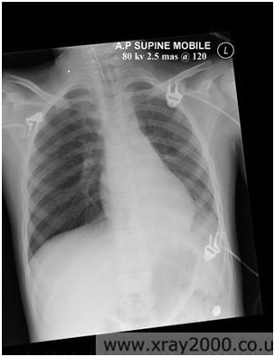
I enjoyed my experience and feel I gained a lot of practical knowledge in patient management. Attached to this piece of writing is an image of a good portable chest x-ray, it highlights the fact that the quality can be diagnostic, however the image is not as good as one performed on a static machine.


Week 8 Year 2

Thursday, February 19th, 2009

Journal

This week on placement I have been at the Royal Victoria Hospital. The hospital is part of the Western General and is dedicated to care of the elderly and an open access service for GP patients and clinics.

I encountered a few challenges throughout the week, these ranged from transferring uncooperative patients and adapting technique to allow for patients conditions. Some patients encountered fear due to suffering pain on movement or weight bearing on fractures. I also encountered many patients with limited or no range of movement. In these instances I had to adapt my technique to obtain the best possible image.

Some patients were a challenge when helping them transfer from their chairs to the table. I found a lot of patients wanted the staff to lift them rather than them transferring themselves on their own.

There was a particular case which I found to be challenging; this involved a patient attending the department from a ward for an ankle x-ray. The patient was brought down to the department in a chair. After confirming all the details I moved the chair to the side of the table. The patient informed me she was frightened to stand, so I advised her to put most of her weight on her good leg while transferring. Getting assistance I assured her we would help her on to her feet and be by her side while transferring her from her chair. On helping her to her feet she seemed to use little effort to support herself making us support most of the weight. After a difficult transfer I then had to try and achieve a good projection. The patient suffered with oedema in her legs and ankles and found it difficult to move them, making positioning difficult.

The AP projection wasn’t too difficult to obtain using the little toe as a marker. However the lateral projection caused a few problems. The patient couldn’t turn onto her side so I then had try and turn her with the use of a pad to hold her in position. It was difficult to see if the patient’s leg was in the correct position due to the swelling but I was able to achieve a good projection.

Week 7 Year 2

Monday, February 16th, 2009

Journal

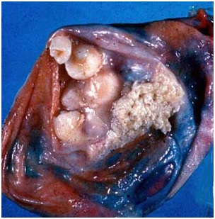
This week on placement I had a number of patients from an oncology clinic for follow up chest x-rays. These patients were all referred due to having conditions called seminoma and teratoma. Both these conditions are cellular cancers which
started, in these cases, in the testes. Seminoma is a radiosensitive malignant neoplasm of the testis, and teratoma is a germ cell tumour composed of multiple cell types derived from one or more of the 3 germ layers.

According to emedicine, 3% of testicular teratomas are known to metastasize in adults and adolescents. There are two types of teratomas, mature and immature. Mature teratomas are usually found in women and are usually found to be benign, whereas the immature teratomas are usually found to be malignant and more commonly found in males. Teratomas are thought to be congenital, but are often not diagnosed until later in life.

The patients had all been referred for follow up chest x-rays by their consultant to check for any metastases in the lungs. I had never heard of any of these diseases before and looking over the request cards I noticed all the patients were all young men, between the ages of twenty five and thirty five. After some research into these diseases I discovered they were both cellular cancers, beginning in the embryonic stage. Once discovered, these diseases are monitored carefully as there is the possibility for them to metastasize.

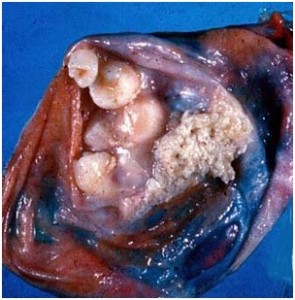
All patients x-rayed had no obvious signs of metastases and were due to see their consultant after their x-ray. Attached to this piece of writing are images and a website I used to research these diseases. Throughout my research I found out both male and females can have this condition, however in this case, all patients were male.

Both seminoma and teratmoas can be found in different parts of the body. According to emedicine, the most common location is sacrococcygeal. As they arise from totipotential cells, they are encountered commonly in the gonads.
The most common location being the ovary, and occurring less frequently in the testes. Occasionally teratomas occur in midline embryonic cell rests and can be mediastial, retroperitoneal, cervical, and intracranial. Cells differentiate along various germ lines, essentially recapitulating any tissue of the body. Examples include hair, teeth, fat, skin, muscle, and endocrine tissue.

Testicular cancer treatment involves radical surgical. This surgery involves the removal of the testicle. As part of the diagnosis of testicular teratoma, tissue diagnosis and distinction from other forms of testicular cancer is important in subsequent management. Regional lymph nodes may also be sampled or removed during surgery. Teratoma of the testicle is relatively resistant to radiotherapy but responds well to chemotherapy as an additional testicular cancer treatment to surgery or as a primary treatment in advanced disease.

Following the surgery or chemotherapy the patient will be closely followed to detect any recurrences early. Follow up includes self-testicular examination, measurement of tumour markers in the blood, serial chest x-ray and regular
abdominal and thoracic CT scanning.


Week 6 Year 2

Friday, February 13th, 2009

Journal

This week I have been on placement in the Western General hospital. This was my first time in this particular hospital so I was naturally a bit apprehensive, not knowing where to go, or knowing any of the staff. However these fears were soon
allayed and, in the end, I really enjoyed it. The staff were lovely, so friendly and helpful and it wasn’t long before I relaxed into my placement. This allowed me to concentrate on familiarising myself with the equipment and finding out how the department runs.

I was curious to see that they performed chest x-rays with the upright bucky. Previously I had only seen a chest x-ray performed with the cassette placed outside the bucky without the use of a grid. I wondered why they chose to perform the examination this way and also how they could be sure they were going to get the entire chest on the image.

After observing this technique a few times, I began to understand the difference between placing the cassette inside or outside of the bucky. The radiographer showed me how the cassette is centred to the bucky, so this gave the radiographer a reference with which to firstly position the patient and then collimate the beam. Once I performed a chest x-ray using this technique I found it quite straight forward.

After performing the examination, I noticed the exposure set on the console seemed very high. When using the bucky for the examination, there is then a need to increase the KVp, which I thought would increase patient dose. I couldn’t understand how this technique could conform to the ‘low as reasonably practicable’ rule regarding radiation, as set out by IRMER 2000 (Ionising Radiation (Medical Exposure) Regulations). However it was explained that this was the radiologists request as this would allow him to see behind the mediastinum and heart for any pathology.

I have since looked at two chest x-rays, one of which I performed using the high KVp and in the bucky, and one taken out of the bucky using the lower KVp from a previous hospital. On comparison, in my opinion, it was possible to see much more detail in the image using the higher KVp and the grid. There is better detail in the lungs and behind the heart as well as through the spine. I found this an interesting method and at some point would like to investigate this method further. I have done some research and since found out the technique used at the hospital can be used to reduce exposure error. An
iontomat is used in collaboration with a high KVp and a grid, while using the smallest possible mAs, therefore reducing patient dose. I have included a link for a website I found while researching the different exposures and techniques,
which gives examples of images at different exposures.

Week 5 Year 2

Saturday, November 22nd, 2008

Journal

This week on placement I performed a number of chest x rays. I felt quite confident at performing these as I had performed a good number throughout each week. However this week was more challenging as I began performing these on patients arriving in the department on trolleys and wheel chairs. Most of these patients were unable to stand for their X ray, meaning we had to adapt the examination to the patient. Observing the radiographer setting up these one after another looked straight forward, however when it came to my turn I was extremely nervous. Several times I asked the radiographer to assist and double check the positioning of the patient and the alignment of the tube.

Once I understood the tube needed to be angled parallel to the cassette it was easier to understand the positioning techniques needed. However I found it difficult to constantly have to examine every individual situation and then try
to evaluate the situation to obtain a good projection.

A routine chest projection is done erect to show any fluid or air levels or possible consolidation. Elderly patients on a trolley, who are very ill or in extreme pain, may create possible problems if they don’t want to sit up or be moved. I found it helped in these situations to take the time to explain to the patients the importance of them sitting up for the projection, as this enables us to achieve a better image for the doctors. It also helped if I reassured them that I would assist them to sit up in their own time. I found when I was nervous in these situations explaining and talking to the patient also gave me
time to calm down and not panic, allowing me time to think about what I needed to do and how I was going to achieve it.

I have learnt numerous things throughout the week, ranging from possible problems that I might face to understanding different situations. One thing I learnt was when patients arrive in the department lying on a trolley with a possible perforation, you need to sit them up and they need to be erect for at least twenty minutes before their examination. Another problem I encountered was a patient who was unable to hold their head up. This meant I had to ask the nurse
who was accompanying them if she minded assisting while I performed the x ray. This required her to be wearing a lead apron while stabilizing the patients the head, as their head could be obscuring part of their chest which could possibly
hide a pathological problem.

I performed a number of (Antero-posterior) AP chests throughout the week, some more challenging than others. However by the end of the week, I found them easier to perform, adapting my technique to a number of what I still thought of
as difficult and challenging situations.

My last challenging situation was on a male patient who had a nasal gastric tube. The request was a chest x ray for positioning of the tube. The challenge with this patient was he had difficulty in standing. I adapted this projection by performing a PA examination, allowing the patient to stay seated in his wheel chair, while taking down the back of the his chair. I was able to get quite a good image, but it only showed the top part of the patient’s stomach. We were able to see the tube on the film but couldn’t see the end. I then asked if it was appropriate to repeat the examination to obtain views of the lower part of the stomach, hopefully allowing us to see the end of the tube. I repeated the examination lowering the cassette and asking the patient to sit upright supporting his self with the top of the cassette. I then coned down to
the appropriate position for the projection and obtained the information needed to show the end of the nasal gastric tube.

?>