Archive for the ‘Reflectives’ Category

Week 9 Mammography Year 3

Thursday, January 14th, 2010

This week I was in the Western General Mammography department. This department has a reputation for being a good department to work in, especially in an observational role, as the staff are highly skilled and very willing to teach.

Prior to attending the department, I had refreshed my memory on the techniques required to these types of examinations, and therefore didn’t have any reservations about what work lay ahead. However, one factor that I was completely unprepared for was the emotional side of type of radiography.

After witnessing my first examination, I quickly realised how difficult breast screenings can be due to a number of factors. The most obvious factor being how personal and intimidating the situation was for the patient. Even I, as the observer, felt that I was intruding in what was a very personal and frightening examination for the patient, and couldn’t help considering how vulnerable they must feel. It was only from my observations and being new in this environment that the impact and nature of this type of examination became evident.

On my first morning the radiographer spent some time showing me around the department before starting the morning list. She proceeded to inform the first patient that she had a student observing in the department and asked her before entering the room if she minded if I observed. The patient didn’t express any issues with this and seemed happy for me to be there and allow the radiographer to explain to me technique and positioning.

Throughout the week I observed many examinations from a number of different areas of referral. The referrals mainly came from one of three areas; new patients from the breast screening unit, ladies who attended the department regularly due to the removal of malignancies and the genetics unit for patients who were in a high risk category due to breast cancer being in the family. I was also able to observe a number of patients attending for stereotactic breast biopsy, ultrasound guided breast biopsy, and core biopsy’s performed in the breast screening unit.

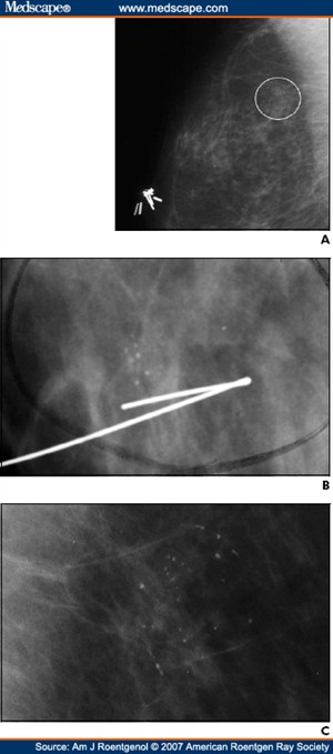
I witnessed a few women with new diagnoses of Ductal Carcinoma in Situ (DCIS). DCIS means that cells inside a patients breast duct have started to turn into cancer cells. With DCIS, these cells are all inside the breast ducts and have not started to spread into the surrounding breast tissue. DCIS is usually impalpable and has different grades from 1 – 3, with 3 being expected to be the most invasive. In the first examination the radiographer performs a stereotactic guidance using one of two biopsy procedures:
§ Core needle (CN) which uses a large hollow needle to remove one sample of breast tissue per insertion.
§ Vacuum-assisted device (VAD) which uses a vacuum powered instrument to collect multiple tissue samples during one needle insertion.
Stereotactic mammography improves accuracy by pinpointing the exact location of a breast mass by the use of a computer and x-rays taken from two different angles. Using these computer coordinates, the radiographer inserts the needle through the skin, advances it into the lesion and removes tissue samples. After confirmation of diagnosis the patient returns to the department for surgical removal of the lesion. Before surgery, the exact location is confirmed by performing another stereotactic procedure called a “localiser” locating the exact position and depth of the tumour. A localising needle is placed near the area of concern and then guided to the cancer with the aid of mammogram or ultrasound. On locating the area they then leave a guide wire in the exact position and depth as a guidance aid for the surgeon. The patient is then able to attend theatre for removal. This type of surgery is classed as Breast-Conserving Surgery and has to be followed up regularly to exclude recurrence.
In my time at the department I was able to observe patients with DCIS not only attend their stereotactic examination, but also proceed to theatre for the removal of the lesion.
Overall it was a very interesting week and I found it quite emotionally intensive. Attached to this piece of writing are images of breast DCIS, which look like tiny grains of salt, with an image of a guide wire in position along with an image of DCIS recurrence.

http://www.breastcancer.org/symptoms/types/dcis/treatment.jsp

This link explains the disease DCIS and the different methods of treatment.

http://cancerhelp.cancerresearchuk.org/type/breast-cancer/about/types/dcis-ductal-carcinoma-in-situ

Explains what the disease is, with a guide to the different grades and treatments and follow up.


Week 8 Elective Placement Year 3

Tuesday, December 22nd, 2009

This week I had been allocated to the rooms that deal specifically with orthopaedic clinics and walk-in GP patients. I have performed many images for the orthopaedic clinics and I feel my ability and skills have grown throughout the week.

There have been audit and review clinics all week and I have had lots of hands on experience. As well as my skills improving I feel I have established a good routine. This is something that I have been working towards for quite a while and it is good to finally have the consistency in my work that a good routine brings. I hope that I will be able to maintain this routine and carry it forward with me on my next placement.

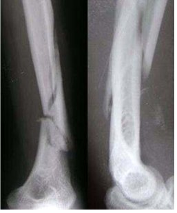
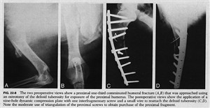
One of the more interesting experiences I have had during this placement involved a patient who had been referred to the department from an orthopaedic clinic. This patient had undergone an internal fixation to her left humerus due to a comminuted fracture 3 weeks previously but the consultant was questioning the alignment of the humerus. A comminuted fracture of a bone is where the fractured bone is in three or more pieces.

The patient was very frightened and was experiencing a large degree of pain. Due to this pain she was unwilling to move her arm. I was able to persuade her by explaining the procedure first and allowing her to manoeuvre her arm by herself; I did however advise her I would help if she needed assistance. She was understandably reluctant to remove her arm from the sling that it was in. I explained I had to take her arm out of the sling to obtain the views needed for her consultant.

To obtain the anterioposterier (AP) view I placed a set of steps with a high support handle attached to them to the left side of the patient, this allowed her to support the weight of her lower arm while she stood in position for the AP view. I explained she would be able to support her arm for the next view by placing her hand on her tummy and holding her arm away from her body. Lifting her arm from the support caused her to cry out in pain. I heard what sounded like a loose screw in the metal and a crepitus sound.

On viewing the images it was clear that 3 of the screws had dislodged from the metal work and away from the bone and the patients arm was not fixed into position. You were also able to view the segments of bone on the images which were in three separate parts. The patient was instructed to return to the orthopaedic clinic for remedial procedures.

Before leaving Perth I managed to follow up on the patient to see if she returned to theatre. The patient had been admitted to one of the wards and was scheduled to return to theatre in the next day.

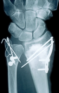
Attached to this piece of writing are images of comminuted humerus fracture and fixations. The first image resembles my patient fractured humerus.


Week 7 Elective Placement Year 3

Tuesday, December 22nd, 2009

This was my first week of a two week elective placement. On arrival to the department I was introduced to staff and shown round by the superintendent. I had been looking forward to this placement and was happy with the schedule that had been arranged for me during my visit. I had specifically requested I would like some hands on with any A & E patients.

It took me a few days to familiarise myself with the systems and equipment, as every room had different types. The department has designated areas for different patients. For my first week I was placed in the rooms which are specifically for A & E and ward patients. I found it all a bit overwhelming at first as the radiographers do almost everything. When a patient arrives from A & E or a ward, the radiographer enters their details and examinations required into the system, after checking their previous history and justifying the examination. They then proceed with the request and then post process the details when finished. Every exposure taken is recorded in detail. They are written on the back of the patients’ request card, the card is then scanned into the system. The exposures are recorded once again during the post processing.
I expected things to be different here but I wasn’t quite prepared for just how different everything was. The equipment here is quite dated such as a Siemens Polydoros 80s which, luckily, I have used previously.

Protocols at the Perth Royal Infirmary are also very different as each orthopaedic surgeon has their own preferences of what views they would like for each examination. So if you have a patient who has had a knee replacement you have to refer to the consultants’ lists and see which views he requests before you perform the examination. Once I understood the procedures and protocols I found this to be a very good system of working.

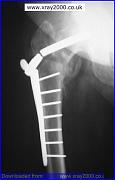
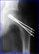
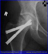
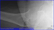
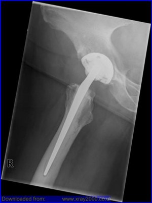
The A&E patient list wasn’t as exciting as I initially thought. However there were lots of post operative patients arriving from the wards and this gave me the opportunity to practice horizontal beam laterals (HBL). In the past I haven’t had much of an opportunity to have hands on practice for this type of examination. It is performed at Queen Margaret Hospital, using the air gap technique with the up-right bucky for trauma patients with a suspected neck of femur fracture. Here they perform their HBL without the air gap technique but the principals are the same. This examination is performed on patients who are post operative, trauma and some orthopaedic clinic reviews. I was given the chance to perform a few and through practice I found them not as difficult as I initially thought.

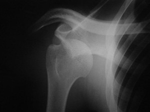
My other experience this week was performing an examination on a patient coming in through A&E with a suspected dislocated shoulder. The examination was quite difficult due to the patients’ pain, although the pain relief began to work and I was able to assist with the views needed. Once the examination was finished I was allowed to return with the patient to the resuscitation unit at A&E and observe while the doctor manipulated the shoulder back into position. It took a doctor and 2 nurses to perform the manipulation. While the doctor was pulling the patients arm down and away from the patients body, one nurse has a bed sheet placed around the top of the patients arm to pull the shoulder up, with the other nurse held the patient on the bed. After a few minutes of pulling, you were able to hear the patients arm slide back into position.

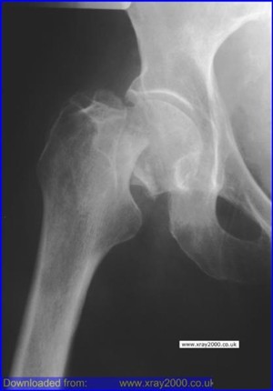
Attached to this piece of writing are images of fractured neck of femur and a hip replacement. A patient with a suspected fracture or a new replacement requires a HBL to show positioning of the fracture or replacement. There are also images of shoulder dislocations.


Week 6 Out of Hours Year 3

Sunday, December 6th, 2009

This week’s placement was my out of hour’s week at Queen Margaret Hospital. I had been really looking forward to this week as I knew there were just two other radiographers working and I was hoping I would have a better chance of being involved with more of the trauma patients arriving in the department. This would give me the opportunity to deal with more challenging situations and the experience of adapting my technique.

Apart from the week having some quiet spells I had quite a few situations to deal with which required me to adapt my technique. I was also able to experience how the department works at night. I found this to be very exciting and I really loved my time in the department. I had advised the radiographers that I had been really looking forward to this week and was hoping to gain as much experience as possible in dealing with any trauma situations.

The first situation I was given was the chance to deal with was an elderly patient with a suspected fractured neck of femur (# NOF). There are a high number of elderly patients attending this department with this type of suspected injury due to the hospital being the main trauma centre. This type of injury has an increasing incidence with age thought to be due to a bone density loss and is more common in elderly females. This is usually due to a lack of oestrogen, which is common due to the menopause. Bone density loss can also been seen in patients taking a variety of medications, such as corticosteroids and thyroxine, with injury mostly due to only minor trauma. Classical features for this type of injury are the patients leg is usually shortened and externally rotated and pain on rotation and tenderness over the femoral neck.

The protocol for views needed at Queen Margaret for trauma situations dealing with suspected #NOF are an anterioposterior (AP) pelvis view and an air gap technique for the lateral (Lat) view. I had previously assisted in this situation before while on placement here, but had never been given the opportunity to perform these views. I had mixed emotions, excited, nervous and very cautious. I was comfortable with the radiographer I was working alongside knowing that they would allow me to proceed with the examination under supervision. I also knew they trusted me not to proceed with the exposure without them checking my positioning was correct first.

The AP pelvis was relatively straight forward although it can be difficult sometimes to see if all the correct anatomy will be on the cassette as patients tend not to lie in the middle of the trolley. I took my time and obtained an AP pelvis with all the relevant anatomy included for a #NOF, which was confirmed on viewing. I then positioned for the horizontal beam lateral (HBL), making sure everything was lined up as I thought it should be and then got the radiographer to check my positioning. I took the exposure and processed the image. On observing the image it was apparent that I could have centred down just a few more centimetres; however, the image showed clearly that the angulation of the head of femur was not in the correct position and the patient’s femur was displaced. We went on to repeat the HBL image which gave me the opportunity to repeat the examination and correct my positioning for a perfect image.

I was also able to visit resus on a few occasions, one occasion being for a gentleman who had fallen down the stairs and had injured has lower right leg. The patient had previous x-rays on his leg as he had a history of Osteomyelitis. This was my first time in resus in a long time so while the doctor was finishing his discussion with the patient the radiographer familiarised me again with the department. I had forgotten that they performed all the examinations with a mobile machine and had been expecting to be using a ceiling mounted tube. Realising this added to my nerves as I immediately thought the examinations were going to be difficult to achieve.

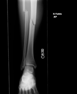
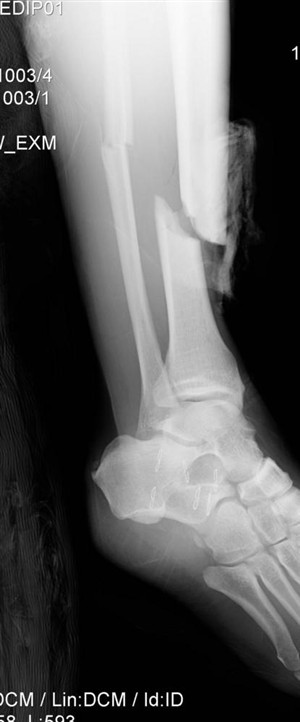
I had observed the patient while I entered the department and had observed that he was covered in blood and his leg was in an unnatural position. I had been advised a fractured tibia is usually associated with a fractured fibula and deformities are common. Deformities or angulation may be obvious on observation due to the foot being abnormally rotated. I obtained the request card to find out what views the doctor was requesting and advised the radiographer what views I thought would be the most appropriate projections.

The doctor had requested an AP and lateral views of the patient’s tibia and fibula from the knee down an AP foot and an AP hand. The most difficult part of the examinations was obtaining the distance for the lateral tib/fib, due to the room size. Moving the patient’s leg for positioning was relatively easy as he had self-anesthetised on alcohol.

I found both examinations very exciting to perform. They were new and challenging situations which I really enjoyed. On the whole the department was quiet due to my shifts being evenings and weekend. I felt I had more of an opportunity to relax and perform more challenging examinations without the pressure of a busy department, although I do feel one week is not enough. I would personally like more of an opportunity to do more out of hours work.

Attached to this piece of writing are images of fractured neck of femur and tib/fib.


Week 5 Endovascular Repair Year 3

Friday, November 27th, 2009

This was week 5 of placement and I was at Queen Margaret Hospital. It was my second day of placement and I was in a newly commissioned digital room. I was excited to be using the new equipment; however I was also very intimidated. Not only was there a new computer to learn to understand, I also found that I had to think differently about each examination. I had only just become comfortable with performing most of the examinations in rooms with a Computed Radiography (CR) system using cassettes, so the difference in environment threw me initially. I found in the beginning I was over thinking things especially when it came to performing extremity work. I was so used to using a cassette and thinking about how the image will look on the cassette and moving it to suit the patients’ position. I found myself thinking about how I could position the patient on the digital system so I could still have a good image.
It settled my nerves to know that even the radiographers I was working with were also a bit nervous using the new equipment. Later that afternoon there was a request of an abdomen examination. I had completed a few abdomen requests that day so didn’t have any problem in performing the examination until, that was, I had read the request in full. The request was for an AP, Lateral and Left and Right Anterior Oblique. I informed the radiographer that I wasn’t confident in performing the Lateral and the Left and Right anterior oblique as I had only read about there positioning techniques but had never saw them being performed.
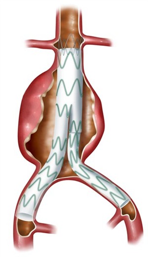
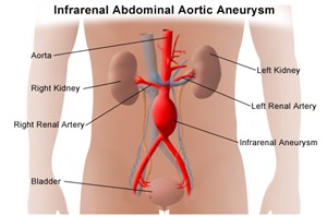
The patient was in for a review due to having an endovascular aneurysm repair. I had never heard of this before and had to ask the radiographer what this was. She explained the patient had a stent placed in his aorta due to an aneurysm which was permanently dilated which is usually caused by a weakening in the vessel wall.
I performed the AP abdomen and the radiographer then performed the others while I observed. The images obtained were great, showing all sides of the stent for the patients’ consultant to review. While observing the images I could see the stent looked like wire mesh which extended from the lower part of the aorta to the upper parts of the common iliac artery, which look like a pair of metal trousers. I found this really interesting and have since done some research into understanding what is involved in endovascular aneurysm repair. I have attached some images and a website address, which shows a good animation of an aneurysm repair, to this piece of writing. This information helped increase my understanding of endovascular aneurysm repair.

http://www.stent-graft.com/

This website gives you lots of information and a good understanding of endovascular aneurysm repair, with animations of the procedure.


My personal development plan year 3

Thursday, October 22nd, 2009

Current Situation

This is my plan of action for my PDP. I aim for development is in A & E radiography. I feel I haven’t had enough practice in adapting my technique in difficult situations. As a student you are not given the opportunity to perform and carry out examination in trauma situations or trolley patients. I feel I have the ability to logically carry out some of the examination I have come across but I very rarely been given the opportunity to carry them out.
I feel the experience in A & E would be of great benefit to me. Giving me the experience and build my confidence when performing them, hopefully making me a better radiographer.

Ideal Situation

My next block for clinical placement I have two ideal situations. First I am in Queen Margaret Hospital for 2 weeks, and 1 of them is my out of hours placement. This hospital is the main hospital for any trauma and I am hoping to be working with someone who will allow me to perform these examinations with supervision.
The following 2 weeks I am on my elective placement at Perth Royal Infirmary. This is a general placement and I have requested time within the department dealing with any trauma and A & E patients. I am hoping to have gained some experience and confidence from my time at Queen Margaret Hospital before proceeding to Perth Royal Infirmary. I am hoping these 2 weeks will be a great experience and of great benefit to me.

Steps to success

  • My 2 weeks placement at Queen Margaret Hospital.
    Learning to adapt my technique and keep calm in difficult situations. While performing as many trauma and trolley examination the 2 weeks I am there.
    • By performing as many examinations as possible on  trauma and trolley patients. by 04 December 2009
  • My 2 weeks at Perth Royal Infirmary.
    • By spending 2 weeks in A & E, and performing as many examinations as possible. Learning the best way to adapt techniques in difficult situations. by 18 December 2009

Overall Completion Date

18 December 2009

SWOT analysis

Strengths

My strength is that I friendly and good at chating to patients in a difficult situations. However this kind of situation is different to what I have ever experienced. I am also entering a new environment in Perth with new staff which I have never met.

Weaknesses

My biggest weakness is my confidence. Even though I know I am capable of most things that are asked of me I still question my ability. I am not sure if this is because we as students don’t get enough clinical time and are not always out on placement. My confidence begins to rise just as we are finishing our clinical placements.

Opportunities

I contacted Perth Royal Infirmary in the hope to get this elective placement. I spoke to a superintendent called Beryl Pritchard and explain what my goals were and what I was hoping to achievement while there.
I have a book which I will be referring to titled, Interpreting Trauma Radiographs. This book covers everything from, Pattern Recognition, Anatomy, Physiology and Pathology of the Skeletal System, Skeletal Trauma, Pelvic Fractures, Chest Trauma, and the Skull and Face.

Threats

Threats to this achievement are radiographers who don’t like students carrying out and performing examinations on patients in a trauma situation or in trolleys.
Another threat is the department is under staffed and I am asked to carry out general examinations to lower patient waiting times while the other staff deal with the trauma patients.

Supporting Resources

Books / journals

Interpreting Trauma Radiographs written by, J McConnell, R Eyres and J Nightingale.

Web links

http://www.wikiradiography.com/page/Pelvic+Trauma+Radiography
http://www.wikiradiography.com/page/Protocol+-+Trauma+Series

Other

Presentation

Reflection

I have just finished a 4 week placement comprising of 2 weeks and Queen Margaret’s Hospital, Dunfermline and 2 weeks at Perth Royal Infirmary and Perth. My experience at Queen Margaret hospital did not give me as much hands on experience as I was hoping for as my out of hours week was quiet. However I did get some opportunities to perform examinations where I had to adapt my technique in order to get good views.

I had the opportunity to practice and obtain a horizontal beam lateral (HBL) on a trauma patient who had a suspected neck of femur fracture. I also had to obtain views on a patient in resuscitation who had fallen down some stairs and fractured his lower leg.

Perth Royal Infirmary (PRI) was completely different from anywhere I had been previously. Being there presented many opportunities to refine and adapt my technique. The Accident and Emergency department is quite small and therefore there are generally not a lot of trauma cases passing through it. There was a big orthopaedic department which allowed me to see and perform techniques I have never observed before. This proved to be a great department to work and learn in, and I feel I have gained knowledge that is invaluable.

On the whole I feel that I have greatly benefitted from this placement. I learnt a large amount regarding adapting my technique for trauma and also views that I have previously read about but have never seen performed before. I think the main thing that I will take away from this placement is the fact that different trusts work in such different ways. I did find the number of changes quite over-whelming initially but by the end of my first week I was starting to adapt to the new routine and understand the system.

I feel writing this PDP has given me an opportunity to evaluate my own capabilities and has also highlighted my fears. By thinking of what aspects of my skill set I want to grow it has allowed me to set a realistic goal for my future development. I do feel this kind of experience is invaluable for me. My confidence did grow by the end of placement however I think this will diminish again the longer I am away from the department.


Week 4 Nuclear Medicine Year 3

Saturday, October 17th, 2009

On week four of clinical placement my work was split between nuclear imaging and Dexa scanning. DEXA stands for dual energy X-ray absorptiometry. It is used to check bone density. Generally, the denser your bones are the stronger they are, and the less likely they are to break.
My time in nuclear imaging was very interesting. For the majority of the time there was only one radiographer, however I feel this was beneficial to my learning as I was able to work extremely closely with her. This was great as she would spend the time to explaining examinations and pathologies in detail to me. The radiographer also reported most of the scans she performed and I would accompany her while she dictated the reports on the day’s scans. She would explain her findings and explain how she had arrived at her conclusions. There were a few scans the radiologist reported which I was able to sit in on while the radiographer and the radiologist reported these.
My time in nuclear imaging greatly increased my knowledge and understanding of the modality. Previously, I was under the misconception that, anyone receiving a radioactive isotope was receiving a really big dose of radiation. I now know this not to be the case. In fact, in the majority of cases the dose a patient receives is usually a lower dose than a normal x-ray. I am not sure why I previously thought that the radiation dose for nuclear imaging was really high; however, I think my misconception is shared by a lot of the general public. Educating the general public about nuclear imaging is maybe an area that the department should look into in order to correct these false assumptions.
On the whole the general public know very little about radiography and I feel all departments should try to educate and inform people about their role in the Allied Health Profession. There were a few occasions while in nuclear imaging when I had to reassure patients that the radiation dose they received was negligible. Further to this, on one occasion, I had to reassure a patient that had incorrectly jumped to the conclusion that they had metastatic bone cancer without any diagnostic evidence. The patient mistakenly presumed that nuclear imaging was used exclusively to find cancer and nothing else.
This lack of understanding or misinformation was also apparent when I worked within the Dexa scanning department. In general I found that the majority of people had never heard of DEXA scanning before and I found myself explaining to friends and family what happened within the department and the reasons we use DEXA scanning. My time in Dexa scanning was very short as I only had about 4 hours there in total. I found this discipline more interesting than I first thought. DEXA scans are used to help find out whether a patient has osteoporosis or possibly at risk of developing it. Most patients who had been referred were conversant with the department as most had a previous history of a fracture. Even with my limited time in this department I gained a lot of knowledge about osteoporosis and bone density.
The radiographer advised me who possibly referrers patients and the different reasons why patients were referred. It was explained how they calculated whether patients were at risk of osteoporosis. This is done by standard deviation, by cross-referencing the patients’ age against an average bone density chart. The graph has been created by scanning hundreds of patients at the age of 35 and recording their average bone density. There are three categories that bone density can be classified as; they are normal, osteopenia and osteoporosis. Osteopenia is the name of the category between normal and osteoporosis. By the end of my time in Dexa the radiographer was allowing me to take some of the patients’ details. I would register patients’ weight and height before positioning them on the table for their scans. In general there are two scans taken, one of the lower spine and one of the hip, two of the main areas at risk from osteoporotic fractures. These are then measured and compared with the osteoporosis risk chart. I found my experience in Dexa scanning very interesting and enjoyable and a very informative and rewarding experience.
Overall my week in Nuclear Medicine and Dexa Scanning has been really enjoyable. Even though both departments were short staffed I was able to have plenty of hands on experience.


Week 3 Year 3

Saturday, October 17th, 2009

My third week of placement was spent at the Victoria Hospital. This was the beginning of my special modalities and I spent 2 days in Magnetic Resonance Imaging (MRI) and 2 days in Computed Tomography (CT).

I did some research before going into the MRI suite hoping to have a better understanding of the modality. Prior to attending my placement, my knowledge of the MRI was only basic. Such as the issues surrounding patient safety, patient positioning, how the machine was constructed and how MRI is best used for soft tissue imaging. However, it soon became apparent, that there was a lot more safety issues than I had previously been aware of. The staff put a large emphasis on the importance of knowing all the safety issues involved when working with an MRI machine with regards to both patients and staff. Considerations have to be given to patients wearing makeup, having tattoos and wearing skin patches. Makeup and tattoos are included in the safety protocol due to some having metallic contents within them and skin patches have to be considered due to the possibility of a metal backing which could heat while in the scanner causing burns to the patient. Also pregnant and breast feeding patient are included in the safety check list due to problems that may arise when administering intravenous contrast agents. I was also surprised to learn that patients having an MRI examination can experience an increase in body temperature due to the radio frequency. The repetitive use of RF pulses deposits energy which in turn causes heating in the patient.

Over my two days in the MRI I was shown how to plan and execute routine examinations. Starting with a localiser scan which was done in a coronal plane and called a scout image. They then proceeded with the different scans available such as axial, sagittal and coronal, and also the angles they use for position slices for different examinations. These are not always obvious, for instance, the angulations for a female pelvis vary from a male due to the fact that the patient has a uterus. If the patient has a uterus the angle is such that the slices are parallel to the endometrial cavity. Other idiosyncrasies include the fact that when a T1 scan is performed the fluid shows up as dark, however, when a T2 scan is performed the fluid shows up bright. Scans are also done removing all the fat from the image; these are called a fat saturation scan.
My next two days were in CT. I found this to be much more stimulating and felt very much more in contact with the patients. It is a very busy department and unfortunately suffering from staffing issues. Because of this, staff was unable to spend time explaining procedures to me, and I was only able to observe. CT imaging, which is also referred to as computed axial tomography (CAT), involves the use of rotating x-ray equipment, combined with a digital computer, to obtain images of the body. Using CT imaging, cross sectional images of body organs and tissues are produced. These images can provide detailed views of soft tissue, bone, muscle, and blood vessels, without sacrificing clarity. I observed quite a few examinations which needed intravenous contrast administered and was able to view examinations with a delayed time sequence for an arterial scan and a portal venous scan.
Overall I enjoyed my time in MRI and CT and hope I have gained a better insight to these modalities. There was an interesting case at the beginning of the week when a patient arrived for an MRI after being referred by an ear, nose and throat (ENT) consultant. The patient had a possible lesion and the ENT consultant wanted to see the position and size. It became clear the lesion had possibly erupted through the skull and embedded itself into the anterior part of the brain. The radiologist referred him immediately for a CT and, unfortunately, this proved to be the case and the patient was referred to the Western General. I would have like to have followed this case up, however again due to the shortness of staff, it wasn’t practical for me to pursue this case any further.

Week 2 Year 3

Saturday, October 3rd, 2009

This week my placement was in the minor injuries department at the Western General. During this week I was aiming to pass my clinical mobile assessment. The minor injuries department deal with the all of the mobile requests for the entire hospital. I was hopeful that I would be provided with enough requests to gain confidence in my work and therefore be competent in performing my assessment.
I encountered a number of difficult situations throughout the week, such as difficulty entering small rooms due to beds, equipment and tables which severely impede the manoeuvring area of the mobile machine. Another problem was the reluctance of some people to move away from an area when a mobile x-ray is being exposed.
The most difficult situation I encountered involved a female patient who required a chest x-ray. The x-ray was due to the fact that she was suffering with progressive shortness of breath (SOB). When we arrived on the ward we identified ourselves to the nursing staff and enquired where to find the patient. The nurse advised us the doctor was still with the patient but we could go in. I could hear the patient who seemed in obvious distress and very agitated. A nurse and doctor were in the room trying everything they could to try and elevate the patient’s condition and help calm her down. However she repeatedly shouted for help stating she just wanted an injection to end her suffering and she wanted her life to be over. The doctor advised her that if she tried to calm down this would help her breathing and she would feel better, she also advised the patient she had phoned for her daughter who she had been asking for, and that she would be arriving shortly. The patient was not responding to the doctor’s efforts to calm her and began to threaten that she was going to jump from her room window.
I found this situation quite distressing and advised the accompanying radiographer that I was unsure how to deal with this from a radiography standpoint. The radiographer was also unsure what to do in this situation and advised the doctor she didn’t feel comfortable performing the request with the patient in the condition she was in. The doctor advised that she needed the x-ray to exclude any underlying conditions and to also plan future treatment. The doctor reassured us that it was in the patients’ best interest to have the x-ray. We duly performed the procedure and, although the patient didn’t refuse the x-ray, she wasn’t very co-operative.
Afterwards, while reflecting on the incident with a radiographer, I wondered what the correct course of action was ethically. In a situation whereby a doctor requests an x-ray in the interests of the patient, however, the patient themselves is in obvious distress about receiving any help or treatments. This seemed like such a grey area. From the patients point of view I can understand if they are in pain and distress and they want to stop the interventions that are keeping them from passing. However, from most patients and relatives point of view, doctors are there to help to alleviate the pain and suffering and must be seen to be doing so. I really felt for this doctor as she was in an awkward situation, for us as radiographers not to perform the x-ray would have made her diagnosis impossible and therefore it would have hindered the patients’ treatment and prolonged her suffering. However, whilst obtaining the projection it felt like we were coercing the patient into something she obviously didn’t want with a complete disregard for her wishes.
Since this experience I have revisited and attached to this piece of writing, The Code of Conduct, as well as The End of Life Strategy document provided by the Society and College of Radiographers (SCoR). Section 3.1in the Code of Conduct states, “Frequently, you may work as part of a multi-professional team and you need to respect the skills, knowledge and contributions of colleagues from other professions and other team members.” While the End of Life Strategy advises, “the SCoR wishes to raise the profile of end of life care within its membership, and to encourage radiographers to consider skills around end of life care as part of continuing professional development”: this document also states, “Radiographers, and imaging and radiotherapy staff in general, need to have a good basic grounding in the principles and practice of end of life care, and many have unmet training needs. They need to have the necessary core competences to enable them to deal with these situations without adverse consequences for the patient, family members and themselves”.

I feel that, after this experience, it is necessary for all radiographers to have situations of this nature, and the appropriate steps to be taken, defined with clear policies and procedures.

Week 1 Year 3

Wednesday, September 23rd, 2009

This was my first week back to University and we went straight out onto clinical placement in the Western General. I had been here on a few occasions so I was familiar with the surroundings and I already knew some of the staff so I was quite excited about being there.

I was working in the main department and I was glad to be back, however I was nervous about going straight back to producing images. Part of me felt worried that it had been so long since I had any practical experience that I may have forgotten everything I had learnt, despite the fact that I had spent time going over books to refresh my memory before I started back. However I do recognise that this was no substitute for hands on experience. The fundamental principles are the same, but there are lots of other factors that are not described in books that only become apparent by doing the job.

My first day went great although, by the end of it, I was exhausted. Being my first day back I was double checking everything and then checking again before I proceeded. While working it was quietly comforting to realise that the knowledge I had gained throughout the previous year’s course was still with me and I was still able to enjoy the job.

Tuesday however wasn’t a good day. The day was going great until late afternoon when I made a very careless mistake. The request was for a chest and femur examination. I was happy performing the examination despite being told that the male patient could possibly be difficult. I proceeded to bring the gentleman into the examination room and after gaining his trust he was happy to co-operate with the procedure. I produced a lovely image of his chest and helped him onto the bed for the femur examination. I had another look at the request form to see which femur was to be examined, put a left marker on the cassette, and discussed with the radiographer that I would proceed to obtain an image from the hip down and then another view from the knee up. The image I produced was extremely clear but I mistakenly x-rayed the right hip. There was no excuse for this mistake and I felt extremely bad for the rest of the day. I obviously went on to obtain a left femur radiograph, but couldn’t shake the guilt of being so stupid.

Under departmental protocol and IRMER regulation 6.8.2 and 6.8.3, patients who undergo a procedure that was not intended, as a result of mistaken identification or other procedural failure, and consequently have been exposed to an ionising radiation dose, should be considered as having received an unintended dose of radiation. My mistake is classed as a radiation incident, and due to this my supervisor then had to report the incident by filling out the appropriate documentation detailing what had happened. Attached to this piece of writing is a copy of the IRMER regulation.

That same afternoon I had another incident that arose from carelessness. I had been working in the same room all day producing good images of chests without any problems. Later that afternoon a projection I performed of a chest had the apices cut off the top. After examining the machine to check my positioning it quickly became apparent how the mistake had arisen. At the Western they perform the chest examinations in the bucky and the person using the room before me had pushed the button on the side of the bucky, moving the positioning of the cassette by accident. This meant that when I had placed the cassette in the bucky, it had not automatically moved up to the correct position for a chest examination.

My mistake was to not check the position of the cassette and become too inattentive. I repeated the examination successfully but the incident taught me a valuable lesson that I was getting in to a bad habit automatically expecting the cassette to be in the correct position. I now make a point to check the position of the cassette every time. The staff were very supportive in the face of my mistakes, reassuring me that they have all made mistakes of their own and reassuring me that I am a good student who produces good work.

I spent that night feeling remorseful about my mistakes and mentally going through the circumstances that led up to the mistake in order to ensure that I didn’t repeat them. The next day I didn’t really want to go back, I really found these mistakes hard to come to terms with, as they were both down to lapses in concentration and therefore preventable.

The rest of the week went well and without incident, it was a hard week, but it is nice to be back.

?>