Archive for the ‘Reflectives’ Category

Week 4 Year 2

Wednesday, November 19th, 2008

Journal

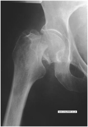
The patient arrived on a trolley in extreme pain, with a suspected fracture to her neck of femur or pelvis. Our initial problem was the patient was lying towards one side of the trolley, with the suspected fracture pressed against the trolley sides. This caused a potential problem as I needed to drop the trolley side in order to position the cassette, however I was very conscious of preventing movement in the injured leg.

The way I overcame this problem was to request assistance from three other members of staff in order to help reposition the patient towards the centre of the trolley. This required us to organise and discuss with the other members of staff the best way to do this, so that everyone knew what was required and moved the patient at the same time. Although the movement caused some discomfort to the patient I ensured that she was fully aware of the reasons behind the move.

Once the patient had been repositioned my next problem was to position the cassette and grid. I am not yet comfortable positioning these as I know a lot of the positioning is done by judgement, usually by drawing an imaginary line with the patient’s position and the cassette from the side and the top of the bed. This helps ensure you have all the appropriate anatomy on the projection. Once I positioned them, I then asked the radiographer to check the position in order for us to gain a good image. After we obtained the image it was clear that the patient had a subcapital fracture of the neck of femur (see attached link).

We then required a lateral image in order to ascertain the extent of the fracture. The protocol for this is horizontal beam lateral (HBL) using an air gap technique. The lateral projection of the hip was done by placing a cassette in the wall stand and not using a grid. Although I didn’t ask why it was obtained without the use of a grid, I think it was done like this to reduce the patients radiation dose by using a low KVp, as a higher KVp would be needed if using the grid. However, when I return in the year I will ask their reasons for using it.

I had seen a few HBL’s using an air gap technique so I understood what was involved, but had never participated before. I placed the cassette in the wall stand out of grid, and then positioned the trolley and patient, placing a 45 degree pad under the sheet, lifting the affected side of the mattress. The trolley was positioned so the neck of femur ran parallel to the cassette. I then lifted the good leg onto the leg box so it was away from the area of interest. We then positioned the tube allowing a horizontal beam which was perpendicular to the cassette. Once I had finished, the radiographer checked
all my positioning before the image was exposed.

On examination of the projections we noticed she also had an exostosis at the top of the femur, which seemed to be coming from the lesser trochanter, I found this very interesting as I had never seen this before.

I now plan to read the radiologist’s report on these’s images to see if he remarks on the exostosis.


Week 3 Year 2

Sunday, November 9th, 2008

Journal

This week on placement I had to perform a chest x-ray on a female patient. The clinical reason on the request card advised the patient had a mastectomy of the right breast 5 years ago and had previously undergone a bone scan showing hot spots with increased uptake of the radioactive substance at certain points. A radionuclide bone scan was performed, this helps show whether a cancer has metastasized to bones, and with the results the consultant had requested the chest x-ray.

The chest x-ray was straight forward; PA erect on the wall bucky, the patient seemed fit with no obvious aches and pains. From reading the request card and before I met the patient, I expected her to possibly be in a lot of pain and possibly a bit weak, however she surprised me, she looked fit, well and happy. I didn’t encounter any problems obtaining this projection as the patient was very slender in build, and I ensured there was no rotation by measuring the distance either side of the patient by slipping my arms between the patients arms and chest and measuring the distance between the cassette and the patients ribs and the distance between the clavicles and the cassette.

This week I have been paying particular attention to critiquing my images and trying to get into a routine. When critiquing my images I start with the patients name and CRIS number, check it’s the correct menu for the image that’s
being performed then going on to check all the appropriate anatomy is on the image and that’s it’s a good diagnostic image with the correct marker on the image. While examining this particular image on the screen it was possible to see a fracture of the 5th rib and a few spots which looked denser than the rest of the bone which I was told may coincide with the hot spot that showed up during the bone scan.

Although I don’t know the full medical history of this patient, she had been sent for a chest x ray after her bone scintigraphy. This is a diagnostic study used to evaluate the distribution of active bone formation in the body, this is done by administering a radioisotope which is used for diagnosis, and to help plan any treatment.

Radioactive isotope can provide diagnostic information about a person’s internal anatomy and the functioning of specific organs.

Reflection

I don’t know much about nuclear medicine yet and haven’t been involved with any patients, however I will spend the time trying to find out a little bit more regarding what is involved when a patient has to undergo a bone scan.

Week 2 Year 2

Sunday, November 2nd, 2008

Journal

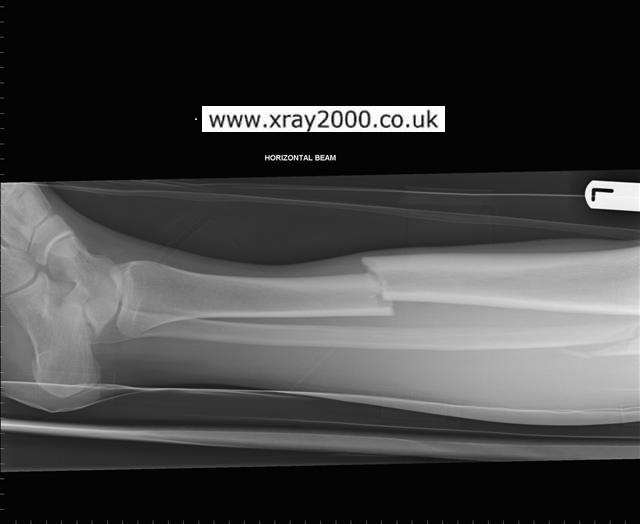
This week on placement I have had a few challenging situations, which I feel I have benefited from. I had a patient who suffers from tremors, so obtaining a good lumbar spine view was more difficult but ultimately successful. However the account I would like to reflect on is my experience with a nine year old patient who had Cerebral palsy. The patient arrived from the orthopaedics clinic with his lower leg in plaster, for a review on a fractured tibia and fibula, querying positioning and alignment of the fracture.

After speaking with his parents and explaining I was a second year student, I asked if they would be happy for me to perform the examination. After being given consent from the little boy’s mother I then explained to her what I
needed to do. The situation was difficult due to the patient being frightened, and strong. His parents had adapted his push chair due to his lower leg being in plaster, and had used a sledge jammed in the chair so their son could sit comfortably. I decided to perform the projections with him sitting in his chair as he was very anxious due to his surroundings and all the new people around him. Once all the appropriate checks were done, i.e. name, address and date of birth, I managed to reassure him as much as possible by spending a bit of time speaking to him and letting him hold my hand, as well as, allowing him touch the cassette to reassure him it wasn’t going to hurt him. This benefited the situation as it calmed his anxiety and he didn’t seem to be as frightened. His mother had to stay with him during the procedures as he kept trying to push the cassettes away, but I was able to get two very good images, with all the detail needed. Although this was not a trauma I had to obtain the lateral projection using a horizontal beam. This situation was a great experience as I had to keep calm in a difficult situation and think outside the box due to the patient’s circumstances.

Reflection

I feel I managed to cope with these situation well. I do feel it was due to being comfortable in my surroundings, due to
working in the department thoughout the summer.

I’m not sure if I would be able to cope so well if I were to be in new or strange surroundings.

I would like to think I would be able to cope in any situation where ever I am, but I have learnt very quickly things are not always that easy.

Theatre Year 2

Friday, October 24th, 2008

Journal

This week on placement I have had a few challenging situations, which I feel I have benefited from. I had a patient who suffers from tremors, so obtaining a good lumbar spine view was more difficult but ultimately successful. However the account I would like to reflect on is my experience with a nine year old patient who had Cerebral palsy. The patient
arrived from the orthopaedics clinic with his lower leg in plaster, for a review on a fractured tibia and fibula, querying positioning and alignment of the fracture.

After speaking with his parents and explaining I was a second year student, I asked if they would be happy for me to perform the examination. After being given consent from the little boy’s mother I then explained to her what I needed to do. The situation was difficult due to the patient being frightened, and strong. His parents had adapted his push chair due to his lower leg being in plaster, and had used a sledge jammed in the chair so their son could sit comfortably. I decided to perform the projections with him sitting in his chair as he was very anxious due to his surroundings and all the new people around him. Once all the appropriate checks were done, i.e. name, address and date of birth, I managed to reassure him as much as possible by spending a bit of time speaking to him and letting him hold my hand, as well as, allowing him touch the cassette to reassure him it wasn’t going to hurt him. This benefited the situation as it calmed his anxiety and he didn’t seem to be as frightened. His mother had to stay with him during the procedures as he kept trying to push the cassettes away, but I was able to get two very good images, with all the detail needed. Although this was not a trauma I had to obtain the lateral projection using a horizontal beam. This situation was a great experience as I had to keep calm in a difficult situation and think outside the box due to the patient’s circumstances.

Reflection

This was a great learning curve for me. Learning not to always judge thing without having any experince. I now know its better to try something and then learn from the experience.

Week 1 Year 2

Friday, October 24th, 2008

Journal

Osteomyelitis

On placement today, I assisted with a patient with severe ostepmyelitis. The patient had previously had his left leg amputated. The patient’s consultant had requested AP and Lateral from knee to foot.

Reflection

I found this a difficult situation, the patient was in extreme pain, with his leg in dressing. The patient was a lovely man, fully aware of the situation and under the circustances, very pleasant and co oprative. Manoeuvring this patient for the exposures we needed was extremely difficult, but it was beneficial for making you think outside the box, to enable you to get the best diagnostic images, without any unnecessary pain for the patient.


Bank Assistant 08

Monday, June 9th, 2008

Working as a Bank Assistant

I have been working as an assistant at the ultrasound clinic with Dr Reid. Observing and assisting with patients who are attending for various clinical investigations.
Listing from, Thyroid investigation, Kidney, uthera and bladder, Liver.
He explains and points out the bile ducts, portal vein and arteries, explains the direction of the beam through the skin, and what is actually being seen. Subcutaneous fat and the colour differences within the tissues. He explained that some peoples liver shows up bright on the ultrasound due to high levels of fatty tissue, where as normally it should show up really dark. The degree of darkness varies as the outline of the kidneys show darker than the centre.

Reflection

I love these clinics, learn something new nearly every week, and enjoy meeting the different patients and finding out about why they have been refered.
Dr Reid makes these clinics enjoyable as well as adding some humor as he has different expressions for different body parts,
ie, gall bladder – tad pole
pancreas – guppy

First every experience in Ultrasound 08

Monday, June 2nd, 2008

Working as a Bank assistant

Dr Reid explained an examination of a thyroid gland.  He explained that there could be one or more nodules/ goiters. He explained multiple nodules doesn’t mean bad news or a problem. Patient can have (hot) meaning a nodule that can cause problems or a (cold) nodule which are usually filled with fluid and not problematic.

Reflection

I found the experience really interesting and informative.

?>