/* Template Name: PageOfPosts */ Angie's CPD » Reflectives

Week 11 Year 3

March 14th, 2010


This week I was in the ultrasound department at the Western General. I had previously worked in an ultrasound department as an assistant so knew what to expect.

The week mainly consisted of obstetrics, and gynecological examination with a little general ultrasound. Through out the week I observed a number of gynecological examinations on women who were perimenopausal and post menopausal. One of the main difficulties I had with these examinations was identifying the ovaries. I generally found it difficult to pick out the ovaries in pre-menopausal women but found it increasingly difficult and sometimes nearly impossible on women who were of peri and post menopausal years. Another issue with this type of exam was that the anatomy changes with women who have had a hysterectomy.

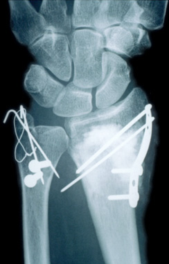
One of the more interesting cases I dealt with was a patient who attended the department for a kidney scan due to a condition called Birt-Hogg-Dube syndrome (BHD). I had never heard of this condition and asked the patient all about it. He went on to tell me he had previously had a pneumothorax and his doctor had heard a crackling in his chest. He was then sent to a cardiologist because they suspected the crackling was coming from his heart. On investigation they could not find anything wrong with his heart and continued with more tests. On further investigation it was found that the patient had tumours in both kidneys, and tests revealed BHD syndrome. This led to him undergoing a partial bi-lateral nephrectomy. The patient was on a follow up appointment checking for any reoccurrence of tumours in his kidneys. This patient had no previous skin lesions and the condition was only identified due to him having a pneumothorax.

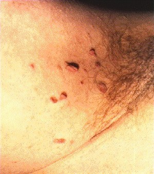
After further research I discovered that Birt-Hogg-Dubé (BHD) syndrome is a hereditary condition that was originally identified as a skin condition by three Canadian doctors. They found certain kinds of skin lesions on the faces and necks of several members of an extended family. In recent years, more symptoms have been linked to this syndrome, namely lung collapses and kidney cancer.

BHD syndrome is caused by mutations in the folliculing gene (FLCN). People who have the mutated gene may have lung cysts or experience collapsed lungs, and may develop kidney cancer. There is no typical BHD patient.

People with BHD syndrome may have none, one, or all of the physical symptoms associated with the condition. The fact that kidney cancer can be one of the symptoms of this syndrome makes it potentially serious. However, only a small percentage of those with BHD actually develop kidney cancer.

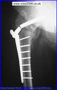
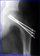
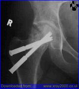
Attached to this piece of writing are images of BHD syndrome.

http://www.nature.com/jid/journal/v128/n1/full/5700959a.html

Article from the journal of investigative dermatology.

BHD syndrome


Week 10 Year 3

March 6th, 2010

This week I was in the Royal Hospital for Sick Kids (RHSC). I had expected this week to be particularly challenging due to the different skills and techniques required while working with children. Being a mother I found myself acutely aware of the thoughts and feelings that some of the children and parents were experiencing.

There were difficulties involved in performing examinations on children while the parents were in the room, especially when the examination required immobilisation and the child was very young. I was constantly aware that I had to display professionalism and confidence in order to gain the trust of the parents, however it was a few days into the week before I was familiar enough with the environment to relax.

Over the week I encountered a number of children with medical conditions that were new to me. One patient I performed a knee examination on suffered from a condition called Dystonia (Sigawa syndrome). This condition is a rare genetic disorder which is characterised by an uncoordinated or clumsy manner of walking and dystonia. Dystonia is a general term for a group of muscle disorders generally characterised by involuntary muscle contractions that force the body into abnormal, sometimes painful, movements and positions.

The request card for this patient advised she had a previous diagnosis of dystonia in her feet, and was now experiencing the inability to straighten her left knee unless she was asleep. I found this difficult to understand how this patient was able to have her knee straight while sleeping but was unable to straighten her knee when she was awake. I have since researched this condition for a better understanding.

Dystonia in Segawa syndrome usually affects the legs; however some children may first develop dystonia in the arms. In some cases, the symptoms of Segawa syndrome may become noticeably worse or more pronounced in the afternoon and evening than in the morning. The symptoms of Segawa syndrome usually become apparent around the age of six years. Children with this condition usually show a dramatic and sustained improvement when treated with a drug called levodopa. Levodopa is an amino acid that is converted to dopamine, a brain chemical that serves as a neurotransmitter. Dopamine is deficient in children with Segawa syndrome. The disorder is caused by mutations of the GCH-1 gene.

The RHSC is also the main centre for Paediatric and Young Adult Spinal Deformity (Scoliosis) in Scotland. During my time there I encountered a number of children attending for a spinal x-rays either pre-operative surgery on their spine or to review their condition to ascertain if there had been any changes in the degree of curvature to their spine. I found performing spinal examinations to be difficult. Mainly due to my lack of experience with this type of examination. I observed and assisted in a number of these examinations and gained a better understanding to what is evolved and the skills needed to produce a good image. It was not always clear by looking at some the children attending for a review x-ray that they had scoliosis until you had x-rayed them. I found it interesting to see how some children manage to compensate their posture to maintain what looks like a normal posture.

Overall my week at RHSC was very interesting and I encountered many new challenges and situations. The staff were very supportive and encouraging which helped to relax me in the new environment. I do feel, however, that I need much more experience in adapting techniques around babies and small children in order to gain confidence in this field. Attached to this piece of writing is an x-ray image of a child with scoliosis, this image is similar to images taken through the week.

http://members.optusnet.com.au/physio/scoliosis.html

A scoliosis is a lateral or sideways curve in the spine that is apparent when viewing the spine from behind.

http://www.nsd.scot.nhs.uk/services/specserv/spinaldeformity.html

Paediatric and Young Adult Spinal Deformity (Scoliosis) Scotland


Week 9 Mammography Year 3

January 14th, 2010

This week I was in the Western General Mammography department. This department has a reputation for being a good department to work in, especially in an observational role, as the staff are highly skilled and very willing to teach.

Prior to attending the department, I had refreshed my memory on the techniques required to these types of examinations, and therefore didn’t have any reservations about what work lay ahead. However, one factor that I was completely unprepared for was the emotional side of type of radiography.

After witnessing my first examination, I quickly realised how difficult breast screenings can be due to a number of factors. The most obvious factor being how personal and intimidating the situation was for the patient. Even I, as the observer, felt that I was intruding in what was a very personal and frightening examination for the patient, and couldn’t help considering how vulnerable they must feel. It was only from my observations and being new in this environment that the impact and nature of this type of examination became evident.

On my first morning the radiographer spent some time showing me around the department before starting the morning list. She proceeded to inform the first patient that she had a student observing in the department and asked her before entering the room if she minded if I observed. The patient didn’t express any issues with this and seemed happy for me to be there and allow the radiographer to explain to me technique and positioning.

Throughout the week I observed many examinations from a number of different areas of referral. The referrals mainly came from one of three areas; new patients from the breast screening unit, ladies who attended the department regularly due to the removal of malignancies and the genetics unit for patients who were in a high risk category due to breast cancer being in the family. I was also able to observe a number of patients attending for stereotactic breast biopsy, ultrasound guided breast biopsy, and core biopsy’s performed in the breast screening unit.

I witnessed a few women with new diagnoses of Ductal Carcinoma in Situ (DCIS). DCIS means that cells inside a patients breast duct have started to turn into cancer cells. With DCIS, these cells are all inside the breast ducts and have not started to spread into the surrounding breast tissue. DCIS is usually impalpable and has different grades from 1 – 3, with 3 being expected to be the most invasive. In the first examination the radiographer performs a stereotactic guidance using one of two biopsy procedures:
§ Core needle (CN) which uses a large hollow needle to remove one sample of breast tissue per insertion.
§ Vacuum-assisted device (VAD) which uses a vacuum powered instrument to collect multiple tissue samples during one needle insertion.
Stereotactic mammography improves accuracy by pinpointing the exact location of a breast mass by the use of a computer and x-rays taken from two different angles. Using these computer coordinates, the radiographer inserts the needle through the skin, advances it into the lesion and removes tissue samples. After confirmation of diagnosis the patient returns to the department for surgical removal of the lesion. Before surgery, the exact location is confirmed by performing another stereotactic procedure called a “localiser” locating the exact position and depth of the tumour. A localising needle is placed near the area of concern and then guided to the cancer with the aid of mammogram or ultrasound. On locating the area they then leave a guide wire in the exact position and depth as a guidance aid for the surgeon. The patient is then able to attend theatre for removal. This type of surgery is classed as Breast-Conserving Surgery and has to be followed up regularly to exclude recurrence.
In my time at the department I was able to observe patients with DCIS not only attend their stereotactic examination, but also proceed to theatre for the removal of the lesion.
Overall it was a very interesting week and I found it quite emotionally intensive. Attached to this piece of writing are images of breast DCIS, which look like tiny grains of salt, with an image of a guide wire in position along with an image of DCIS recurrence.

http://www.breastcancer.org/symptoms/types/dcis/treatment.jsp

This link explains the disease DCIS and the different methods of treatment.

http://cancerhelp.cancerresearchuk.org/type/breast-cancer/about/types/dcis-ductal-carcinoma-in-situ

Explains what the disease is, with a guide to the different grades and treatments and follow up.


Week 8 Elective Placement Year 3

December 22nd, 2009

This week I had been allocated to the rooms that deal specifically with orthopaedic clinics and walk-in GP patients. I have performed many images for the orthopaedic clinics and I feel my ability and skills have grown throughout the week.

There have been audit and review clinics all week and I have had lots of hands on experience. As well as my skills improving I feel I have established a good routine. This is something that I have been working towards for quite a while and it is good to finally have the consistency in my work that a good routine brings. I hope that I will be able to maintain this routine and carry it forward with me on my next placement.

One of the more interesting experiences I have had during this placement involved a patient who had been referred to the department from an orthopaedic clinic. This patient had undergone an internal fixation to her left humerus due to a comminuted fracture 3 weeks previously but the consultant was questioning the alignment of the humerus. A comminuted fracture of a bone is where the fractured bone is in three or more pieces.

The patient was very frightened and was experiencing a large degree of pain. Due to this pain she was unwilling to move her arm. I was able to persuade her by explaining the procedure first and allowing her to manoeuvre her arm by herself; I did however advise her I would help if she needed assistance. She was understandably reluctant to remove her arm from the sling that it was in. I explained I had to take her arm out of the sling to obtain the views needed for her consultant.

To obtain the anterioposterier (AP) view I placed a set of steps with a high support handle attached to them to the left side of the patient, this allowed her to support the weight of her lower arm while she stood in position for the AP view. I explained she would be able to support her arm for the next view by placing her hand on her tummy and holding her arm away from her body. Lifting her arm from the support caused her to cry out in pain. I heard what sounded like a loose screw in the metal and a crepitus sound.

On viewing the images it was clear that 3 of the screws had dislodged from the metal work and away from the bone and the patients arm was not fixed into position. You were also able to view the segments of bone on the images which were in three separate parts. The patient was instructed to return to the orthopaedic clinic for remedial procedures.

Before leaving Perth I managed to follow up on the patient to see if she returned to theatre. The patient had been admitted to one of the wards and was scheduled to return to theatre in the next day.

Attached to this piece of writing are images of comminuted humerus fracture and fixations. The first image resembles my patient fractured humerus.


?>