Archive for the ‘Reflectives’ Category

e-KSF Personal Development Review

Sunday, February 17th, 2013

Gateway

Year 1 e-KSF   link to evidence

No.  Dimension Name  Dimension Type Foundation Outline Level Full Outline Level Current Level  Evidence
C1  COMMUNICATION Work Related 2 2
C2  PERSONAL AND PEOPLE DEVELOPMENT Work Related 1 2
C3  HEALTH, SAFETY AND SECURITY Work Related 1 2
C4  SERVICE IMPROVEMENT Work Related 1 1
C5  QUALITY Work Related 1 1
C6  EQUALITY AND DIVERSITY Work Related 1 2
HWB3  PROTECTION OF HEALTH AND WELLBEING Work Related 1 1
HWB6  ASSESSMENT AND TREATMENT PLANNING Work Related 1 3
IK1  INFORMATION PROCESSING Work Related 1 1

PDP Year 4

Friday, April 1st, 2011

Current Situation

The beginning of semester 2 of 4th year. This is my plan of action for my PDP. My aim for development is to continue to get as much hands on experience within the general department as possible. I still feel I haven’t had enough practice in adapting my technique in difficult situations. Although I had more of an opportunity in the last clinical block, I still feel I need more. There was one very busy week with very little staff on my last clinical block and I was allowed to carry out most examinations on my own without any interventions from other staff. This allowed me to perform examinations without feeling intimidated or nervous and this helped to increase my confidence.
However there are still examinations that I shy away from because I feel I am unable to perform them as well as some other examinations but this is an area that I am determined to work on.

Ideal Situation

An ideal situation would be to have the same opportunity again, especially because I have my fit to practice very soon. Confidence is a big problem for me and when I am working along side a member of staff that doesn’t know me I know it is going to take time before they allow me to work on my own. I like to perform the complete examination, from checking the patients previous history, to cleaning the room at the end. I feel this allows me to get into a routine knowing everything has been done. I enjoy working with others and completely understand when someone is helping me, however because our clinical placement are in blocks and you continually work with different staff members, I feel establishing a routine and being allowed to continually perform it helps to cement good practice and remove the possibility of errors.
My action plan for next block is to establish and maintain a routine that works for me and to tackle projections that I am not comfortable with, mainly axial shoulders, lateral scapulas and horizontal beam hips. I continually get told I am a good radiographer however until I feel I can perform to the level I think is acceptable then my confidence will always be low.

Steps to success

  • Continue to have as much hands on within the general department.
  • Tackle all examination that I am not confident in performing to help increase my performance and confidence.
  • Perform more examinations that allows more hands on which allows me to adapt techniques.

Overall Completion Date

01 April 2011

SWOT analysis

Strengths

My strength is that I love the job and I strive to be as good as I can be. I as love the interaction with the patients and I am friendly and good at chatting to patients in a difficult situations.

Weaknesses

My biggest weakness is my confidence. Even though I know I am capable of most things that are asked of me I still question my ability. I know this will increase over time however with the little clinical time we receive it is difficult as there is never enough time to perform everything you would like to. My confidence begins to rise just as we are finishing our clinical placements.

Opportunities

On my last block I am at Queen Margaret Hospital. I would like to use this week to work alongside a member of staff who will advise and supervise me while I am performing the examinations I don’t feel comfortable with, mainly axial shoulders, lateral scapulas and horizontal beam hips, so I can overcome my nerves when performing them.

Threats

Threats to this situation can be working alongside a member of staff who interacts when you are performing an examination. This could be them checking your positioning of the patient. Some staff move the patient from there original position and then put them back to exactly the same position you had them in. Some radiographers do that through habit and are aware of it and apologise, however some do it all the time and don’t acknowledge they have done the exact same thing as you had done and this can be frustrating as a student. It also knocks your confidence and you end up questioning your ability, especially if your working alongside someone that does it continually.

Supporting Resources

Books / journals

Carver and Carver.
Clarks.

Web links

http://www.wikiradiography.com
http://www.e-radiography.net

Electronic resources

Presentations

Reflection

I have just finished my very last placement as a student. On reflection I feel I have had a very productive year. I also feel my confidence has increased dramatically over this last year, allowing me to gain the confidence to perform my “confidence to practice” in an out of hours placement. I really enjoy tackling the challenges of A & E situations as well as the challenges required for dealing with trauma patients which require me to think about the technicalities required when performing some examinations.
I also like the fact that as my confidence increases I relax and enjoy the job more. I do release I have so much more to learn and I look forward to doing this in the future.

Colonic Stenting 29/03/11 Year 4

Tuesday, March 29th, 2011

Description

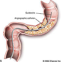
Schematic drawing of a tumorous lesion in the rectosigmoid junction traversed with a guidewire and catheter. C, Terumo guidewire (left) and angiographic catheters (right), Cobra and Headhunter types. D, Schematic drawing of the stent instrument advanced over the superstiff wire and the released stent in place.

Imaging consult colonic stenting.jpg


Week 13 Year 4

Monday, March 21st, 2011

21 March 2011

This was my final week of placement as a student. It was quite daunting as I was very conscious of the fact that the next time I work in a radiography department I will be a qualified radiographer. It was also very exciting as, after 5 years, I can now see the end of the course in sight.
During the week I carried out a general practitioner (GP) request on a female patient who had presented to her GP with pain and tenderness in her forearm. The clinical history was brief so I asked the patient for more details. She showed the area of the pain which looked a little swollen but nothing remarkable. She informed me there had been no trauma and she didn’t understand why it was painful. I then continued with the examination of an AP and lateral forearm.
On processing the image it was very clear there was something obviously amiss with the patients’ ulnar. It looked as if something had taken a clean bite out of the patients’ bone, I had seen a similar case previously and thought it looked like osteosarcoma. I asked another radiographer for her opinion and we decided to get a radiologist to have a look before letting the patient leave. It was the opinion of the radiologist that the patient did have osteosarcoma and he was going to telephone her GP straight away so she could get an urgent referral to the osteosarcoma team.
I found the patients symptoms for this very surprising and I could understand why it took her so long to visit her doctor. The patient was 32 years of age with an 18 month old baby so it was understandable that she first thought she may have bumped it without realising and it would get better.
Osteosarcoma is a primary malignant tumour of bone and the cause of it is unknown. According to Shenoy at el (2007) osteosarcoma is the most common malignant bone tumour which conflicts with the opinion of Pretorius and Jeffrey (2006) who state osteosarcoma as being the second most common primary bone tumour after multiple myeloma. It is also the opinion of Pretorius and Jeffrey (2006) that sarcomas tend to undergo hematogenous spread, with pulmonary metastases being the most common.
According to Wang and Houston (2005) most osteosarcomas are sporadic, whereas inherited predisposition accounts for a small number of cases, along with this 20% of patients with osteosarcoma present clinically with overt metastatic disease, with the presence of metastatic disease being a strong predictor of a poor outcome. The disease is believed to originate from primitive mesenchymal cells but also may arise from pluripotential mesenchymal cells. Osteosarcoma is a deadly cancer and is commonly fatal when it metastasizes to the lungs. It usually forms as a single lesion on long bones, with the distal femur being the most common, then the proximal humerus and proximal tibia.
Several tests are necessary to diagnose osteosarcoma. Initially, a blood test followed by imaging tests to detect any other tumours and their location within the body. This can be performed by an X-ray, MRI, CT scan or bone scan. Confirmation of osteosarcoma can also be done by performing a biopsy to remove a piece of bone tissue.
Treatment for osteosarcoma depends on the location of the tumour and the severity of the illness. Treatments may include neoadjuvant therapy which is the administration of a therapeutic agent before the main treatment of chemotherapy to destroy cancerous cells and prevent the growth of new malignant cells. The aim of neoadjuvant therapy is to reduce the size or extent of the cancer before using radical treatment intervention, thus making procedures easier, more likely to succeed, and reducing the consequences of a more extensive treatment technique that would be required if the tumour wasn’t reduced in size or extent. Additionally, radiation therapy, which uses high-energy radiation beams to shrink tumours and destroy cancer cells may also be used to treat osteosarcoma. In advanced stages of osteosarcoma, doctors may suggest amputation to remove the affected limb. Advances in surgical techniques have allowed a reduction in the number of amputations associated with osteosarcoma. Tumours can now be removed from the affected bone without removing a limb, this is done by using artificial bones or bones from other parts of the body. These advances in surgical techniques to treat osteosarcoma have resulted in higher survival rates, and high limb salvage rates associated with this type of cancer. Attached to this writing are images taken from x-ray 2000 and the childrens specialist website.

Pretorius, E. S. and Jeffrey A. S. 2006. Radiology secrets. 2nd ed. Philadelphia: Mosby Elsevier

Shenoy, R. Pillai, A. Sokhi, K. Porter, D. Ried, R. 2008. Survival trends in osteosarcoma of humerus. European Journal of Cancer Care 17, pp. 261–269.

Wang, L. 2005. Biology of Osteogenic Sarcoma. Cancer Journal. pp. 294-305.

Ehow Health. 2011. How long are treatments for osteosarcoma. [online] Available at: http://www.ehow.com/how-does_5251031_long-treatments-osteosarcoma_.html [Accessed March 27]

Childrens Specialists. 2011. Musculoskeletal Tumors and Infections. [online] Available at: http://www.cssd.us/body.cfm?id=1238 [Accessed March 27]

Week 12 Year 4

Monday, March 21st, 2011

This week I was working in the interventional room at the Western General Hospital. There were a number of procedures that were new to me, so it was a very interesting week. Some of the procedures that I assisted with were barium enemas, water soluble enemas, Hickman lines, colonic stenting and gastrostomy tubes. There were a number of Hickman line procedures where I was allowed to perform the screening and also carry out nursing duties. All the patients I assisted with were all receiving their Hickman lines for treatment of different types of cancers.
Hickman lines consist of a soft plastic tube that is tunneled beneath the skin and placed in a large vein. The insertion is carried out for a number of reasons and depending on the requirement of the line will depend on the number of connectors required, which can be single, double or triple lumen catheter. These catheters can then be used to give fluids, drugs and for taking blood samples. This procedure is usually carried out to save the patient repeatedly having to endure needles for giving or providing blood samples or for administering drugs.
The radiologist marked the vein using ultrasound then proceeded to administer a local anaesthetic. He then made two cuts in the patients’ chest, one to tunnel the catheter and the other near the collarbone. The line was then inserted into position with the use of fluoroscopy screening, this allows real time visualisation of the line and the anatomical structure ensuring the line is positioned correctly. Once the Radiologist was happy with the position he then flushed the line to ensure it was working correctly. He then inserted two stitches at either end of the tube insertions to secure the catheter in position and to stop it from moving. Patients will have their Hickman lines in situ for the period of their treatment before having them removed. The use of fluoroscopy substantially decreases the amount of radiation needed to produce clinically useful images (Pretorius and Solomon 2006).

Another procedure that I assisted with was a colonic stenting on an elderly female patient with an abdominal obstruction. The patient had previously been diagnosed with colon cancer and had a 10 centimetre stricture causing an obstruction. The patient had previously been offered surgery to remove the tumour but had refused it as she didn’t want to undergo a major operation.
The procedure was carried out by a radiologist and a gastroenterologist consultant. The gastroenterologist consultant proceeded to guide the colonoscopy into the patients’ colon to try and pass the stricture. The radiologist then proceeded to insert a guide wire beyond the blockage, using fluoroscopy guidance. A small catheter was then positioned over the wire and the guide wire removed. Another wire was then put down the catheter with a balloon and stent on the end which was ready to be expanded when it was in the correct position. Contrast dye was given to show the bowel outline and the exact position of the blockage for positioning of the stent. Once in the correct position the wire was removed leaving the stent in position. According to Dionig at el, (2007) management of colorectal obstruction by using a metallic stent is a safe and effective procedure with good technical and clinical success; the use of stent can prevent the need for surgery in patients with disseminated disease; it can prevent both temporary and permanent stomas and may mitigate the need for emergency operations for colonic obstruction.
This procedure proved to be a difficult procedure as it was difficult for the gastroenterologist to pass the strictures part of the bowel with the procedure taking longer than hoped.
Possible complications with this procedure can be the movement of the stent where it may migrate further down the bowel. This will then cause the stricture to return and result in the patient having to endure removal of the stent with a further second stent having to be positioned. Another complication can be the risk of perforation to the bowel wall.

Pretorius, E. S. and Jeffrey A. S. 2006. Radiology secrets. 2nd ed. Philadelphia: Mosby Elsevier

Dionigi, G. Villa, F. Rovera, F. Boni, L. Carrafiello, G. Annoni, M. Castano, P. Bianchi, V. Mangini, M. Recaldini, C. Lagana, D. Bacuzzi, A. Dionigi, R. 2007. Colonic stenting for malignant disease: Review of literature. Surgical Oncology, 16 pp. 153-155.

Week 11 Year 4

Monday, March 14th, 2011

13 March 2011

This week I was in the Royal Hospital for Sick Kids. It was a very interesting week with me experiencing and learning a lot. Paediatric radiography is very different to general adult radiography. Rules and protocols are strictly defined for paediatric radiography and students who are on placement are made aware of them very quickly.
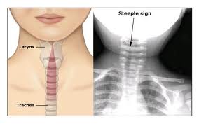
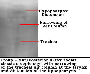
While there I was able to attend the weekly A & E meeting. This meeting allows the A & E consultants and the radiologists to discuss any interesting cases or cases where there are questions surrounding patients’ diagnosis or follow up. Being able to sit in on these meetings, from a student’s point of view, was extremely interesting and very useful. One patient on the list of discussion was a two year old who had ingested a liquid tab designed for washing machines. What made this particularly interesting was a feature on the x-ray that was pointed out and referred to as a steeple sign. They explained this sign is usual in children with croup or children who have experienced near drowning and referred to it as being laryngotracheobronchitis. This steeple sign is a tapering of the trachea which can be seen superiorly on a frontal chest radiograph and is reminiscient of a church steeple.
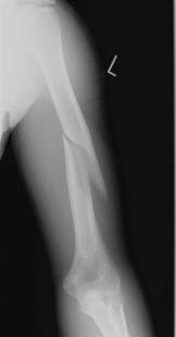
Another case that was discussed was regarding a possible non accidental injury (NAI) of a 7 month old child who had been brought in by his parents due to continued crying and seemed to show signs of a sore arm. It was discovered on x-ray the patient had a spiral fracture of the humerus with minimal displacement. However the fracture travelled from the distal humerus and ran over half way up the shaft of the humerus. It was highlighted by the consultant at the meeting as the patient had been seen previously with a case of severe scabies and also the patient had multiple bruises along with a number of other suspicions which were not discussed. The consultant also questioned the mechanism of injury and said the story did not seem to relate to the extent of the injury. He went on to inform the radiologist that he had reported the case to the child protection team for further investigation.
Later on in the week I was in ITU performing a chest x-ray on an 18 month old patient. The little girl suffered from a rare condition called Jeune syndrome and had previously undergone surgery at Great Ormond Street Hospital to reduce the restriction of her breathing due to her ribs not growing and crushing her lungs. The consultant had informed us that he was extremely concerned for the child and he was unsure if she was going to recover. She had now had a tracheostomy performed to try and assist with her breathing however this didn’t seem to be helping.
An article from emedicine refers to Jeune syndrome as a disorder of bone growth caused by changes in the IFT80 gene. Common signs include a small chest and short ribs which restrict the growth and expansion of the lungs often causing life threatening complications. Other symptoms can include shortened bones in the arms and legs, unusually shaped pelvic bones, and extra fingers and/or toes (polydactyl). Children that survive the difficulties of the breathing and lung challenges at infancy, can then go on to later develop life-threatening kidney problems or heart defects and a narrowing of the airway. Less common features of Jeune syndrome can include liver disease, pancreatic cysts, dental abnormalities, and an eye disease called retinal dystrophy that can lead to vision loss.

Attached to this writing are images on the various topics discussed above.

Children’s Specialists. 2011. Musculoskeletal Tumors and Infections. [online] Available at: http://www.cssd.us/body.cfm?id=1238 [Accessed March 27]

Aswcpoets. 2011. Steeple sign. [online] Available at:http://www.aswcpoets.com/wp-content/uploads/2011/01/croup_steeple_sign.jpg [Accessed March 12]

Radiopaedia. 2011. Croup – steeple sign. [online] Available at:http://radiopaedia.org/cases/croup-steeple-sign [Accessed March 12]

Radiopaedia. 2011. Skeletal dysplasia. [online] Available at:http://radiopaedia.org/articles/skeletal-dysplasia [Accessed March 12]

Dukes Orthopaedics. 2011. Pediatric Supracondylar Fractures of the Humerus [online] Available at: http://www.wheelessonline.com/ortho/pediatric_supracondylar_fractures_of_the_humerus [Accessed March 12]

Emedicine. 2011. Genetics of Asphyxiating Thoracic Dystrophy (Jeune Syndrome)
[online] Available at:http://emedicine.medscape.com/article/945537-overview [Accessed March 12]


Week 10 Year 4

Sunday, March 13th, 2011

This week I was on an out of hour’s placement at Queen Margaret hospital. It was my very last block of placement as a student and I was preparing for my competence to practice. I had mixed feelings of real excitement coupled with extreme nervousness. Despite the fact that I have worked at Queen Margaret many times, the pressure was greater this time as I really wanted to perform to an exceptionally high standard. The first day went well however there was the usual period of trying to familiarise myself again with the computerised radiology information system (CRIS).
I had informed the radiographer that this was the last opportunity for me to get as much hands on as possible before my competency to practice and therefore I asked her if I could perform as many of the examinations as possible and be treated as if I were getting assessed. There was an orthopaedic clinic on until 7.30pm during which I managed to get lots of hands on experience albeit with relatively straight forward examinations. However after the clinic finished the requests were predominately A&E referrals, along with ward and mobile requests. One of the exams I performed was a mobile chest examination on a 73 year old female with reduced air entry and basal crackles. On entry to the ward I identified myself as the radiographer to the duty nurse and received the request card. I then performed the usual ID checks with the patient and prepared myself along with the equipment for the examination.
Once I had everything in place I proceeded to ask the patient who was sitting on the edge of her bed if she would get up onto her bed so I could perform the examination. Chest x-rays are usually performed posteroanterior (PA) as this reduces magnification of the heart; however mobile requests or ward patients that are unable to attend the department are usually performed anteroposterior (AP). According to Clarks (2005) ward radiography is normally complicated by a variety of situations, some of these complications being the patients’ condition, degree of consciousness and cooperation.
Most books demonstrate mobile x-ray examinations being performed with the patient in an AP position. According to Ohioswallow an AP portable chest is inferior to a PA or lateral. Problems with an AP chest include magnification of the heart shadow, artifacts from lead wires, lines, bedsheets, and skin folds, patient rotation, visualisation of the chest in one plane only and variable exposure factors related to the equipment used.
The radiographer who was with me, then stopped me and advised me to just leave the patient where she was sitting and to perform the examination with the patient sitting on the edge of her bed. I had not seen this done and couldn’t understand how I was going to position the cassette.
I told her that I had once performed a chest examination with the patient sitting on the edge of a trolley using the upright bucky but although I understood the theory of what she was asking I was not sure of the best way to position the cassette in this situation. She then advised me to position the cassette on the patients lap and ask her to give it a cuddle placing her finger tips under the bottom. After following her instructions I was able to perform the examination without having to move the patient at all.
This was one of those occasions that allowed me to build on my knowledge and gain a better understanding from a very experienced radiographer. I was able to perform the chest x-ray without causing upset or difficulty to the patient as well as reducing positioning problems for myself, while achieving a diagnostic PA image.
I felt this was a much easier technique to achieve a diagnostic image and is definitely one technique that I will use in the future.
As a student you find yourself working with many different radiographers and quite often they’ll have their own way of working and performing examinations. This has both advantages and disadvantages. The main advantage is that you get shown many different ways of performing the same examination and an insight into why they prefer that technique. As a student you are then able to try the different techniques you have been shown and choose which one you find the most suitable. Conversely, many varied techniques can be a disadvantage if you already have your own style of performing an examination only to be shown other techniques and potentially get confused as to the best method. Working with this particular radiographer definitely had its advantages for me and I feel that I finished the week a better radiographer than when I started.

Clark, K.C. 2005. Clarks positioning in radiography. 12th ed. London: Arnold.

http://www.ohioswallow.com/extras/0821417088_chapter_05.pdf

Week 9 Year 4

Sunday, January 9th, 2011

09 January 2011

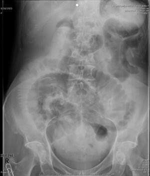
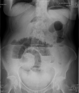
While on placement this week we had a patient arrive via Accident and Emergency (A&E) with a suspected abdominal obstruction. An abdominal obstruction is where there is a suspected obstruction in the small or large bowel which has become impacted. Patients being referred for suspected abdominal obstruction have to be in an upright position. The contents of the intestine and movement and motion can come to complete arrest. Symptoms usually include cramping pain, vomiting, obstinate constipation, and lack of flatus. Clinical diagnosis is confirmed through an abdominal X-ray. After carrying out the abdominal x-ray on this particular patient it was evident she had something unusual going on as the centralised part of her small bowel was extremely dilated.
Common causes of abdominal obstruction can be adhesions which usually develop after some form of pelvic surgery, hernias, and tumors. Other causes are diverticulitis, foreign bodies such as gallstones, and volvulus (twisting of bowel on its mesentery). However specific segments of the intestine are affected differently.
In a simple abdominal obstruction, a blockage can occur without any compromise to the vascular supply. All foods, fluids and digestive secretions ingested after an obstruction has occurred along with any gas accumulate above the obstruction. This causes the proximal bowel to distend, and the distal segment to collapse. This then causes the normal process of absorption and secretions of the mucosa to become depressed and congested. The bowel walls then reach deluge due to an excessive accumulation of serous fluid in the tissue spaces in the body cavity. The abdominal cavity then progresses to distend, causing strengthened peristalsis (contraction and relaxation of the alimentary canal) and an upset of the normal condition and functioning of secretions. This then increases the risks of dehydration and the advancement to a strangulating obstruction.
Strangulation of the obstructed bowel causes a compromised blood flow and can occur in a significant number of patients with small bowel obstructions. A strangulated obstruction can progress to infarction (an area of tissue death (necrosis) due to a lack of oxygen caused by obstruction) and gangrene in a short period of time.
Venous obstruction occurs first, followed by arterial occlusion (closure or blockage of a blood vessel), resulting in rapid ischemia. The ischemic bowel becomes a deluge with edema and infarcts (areas of tissue death), leading to gangrene and perforation.

Perforation can occur in an ischemic section (typically small bowel) or when there is a large amount of dilation. Perforation risk increases when the cecum is dilated to a diameter equal to or greater than 13 cm.
Symptoms and signs from an obstruction of the small bowel are usually;
• abdominal cramps around the umbilicus,
• vomiting
• partial obstruction may develop diarrhea.
• absence of strangulation, the abdomen is not tender.
• Hyperactive, high-pitched peristalsis with rushes coinciding with cramps
• dilated loops of bowel maybe palpable
• infarction, the abdomen becomes tender and auscultation reveals a silent abdomen or minimal peristalsis.
• Shock and oliguria (a low output of urine) indicate late simple obstruction or strangulation.
Symptoms and signs of a large bowel obstruction are milder symptoms. These develop gradually differing from small-bowel obstruction.
• Increasing constipation leads to obstinate constipation and distention of the abdomen
• Vomiting may occur
• Lower abdominal cramps and non-production of feces
• Examination usually shows a distended abdomen with gurgling sound caused by the movement of gas in the intestines (borborygmi).
• no tenderness, and an empty rectum
• mass corresponding to the site of an obstructing tumor may be palpable.
• Systemic symptoms are usually mild, with fluid and electrolyte deficits being uncommon.

Care for patients with small and large bowel obstruction is similar. Treatment used for an obstructed bowel can be, Nasogastric suction, IV fluids and IV antibiotics if bowel ischemia suspected. Sometimes specific measures of resection are need in adults with an obstruction of the duodenum.
If the obstruction is caused by diverticulitis a perforation is often present. If a perforation is evident then removal of the section involved is required. Resection and colostomy are then carried out, and the joining of blood vessels (anastomosis) is postponed.

Carver, E. and Carver, B. 2006. Medical imaging: techniques, reflection and evaluation. Edinburgh: Churchill Livingston Elsevier.

http://digestive.niddk.nih.gov/ddiseases/pubs/intestinaladhesions/AbdominalAdhesions.pdf

http://www.merckmanuals.com/professional/sec02/ch011/ch011h.html#BGBBGBGG


Week 8 Year 4

Saturday, January 8th, 2011

Week 8

This week I was out of hours. It was a quiet week; however there were a few mobile exams and a few occasions where I had to adapt my technique in order to achieve a good result. A few of these examinations being chest x-rays on patients who were unable to co-operate due to either being unconscious or medicated. I also had a few wrist x-rays pre and post manipulation where I had to adapt my technique to perform them.
Patient Positioning
The optimal positioning for obtaining a chest radiography is with the patient postero-anterior (PA) and erect (Clarks 2005). However this is not always possible, due to patient conditions some radiographs have to be obtained antero-posterior (AP) with the patient in a sitting or semi-recumbent or supine position. Patient positioning has a significant influence in the appearance of air or fluid, and blood vessels within the chest: Air usually rises to the highest point within the chest cavity. Pathologies such as a pneumothorax are mostly seen in the apex of the lung in an erect chest x-ray. When the patient is supine, the highest point in the chest lies alongside the heart and mediastinum. According to medscape 2010, a pneumothorax may cause increased lucency adjacent to these structures, which appear to have a better-defined outline than normal, often with no lung edge visible.
On an erect film any fluid lying in the lungs usually collects at the lung bases and appears as cloudy or opaque and obscures adjacent structures. Fluid levels in the lung are higher on the lateral wall of a chest image and lower at the mediastinum. When performing a supine examination, any fluid in the lungs would lie along the posterior chest wall.
A technical problem encountered by student radiographers while performing mobile chest x-rays are Lordotic AP films sometimes referred to as an apical lordotic view.  A lordotic image is produced when the tube angle is not in the correct position. One of the first things you are taught when performing mobile chest exams is to always try to sit the patient as up-right as possible while giving consideration to the patients condition. Although you are taught in lectures about how a lordotic image is achieved, it sounds easy to avoid.  This has been something I have had to consider a lot on placement this week.
A technique shown to me while on placement which should eliminate the chance of producing a lordotic image is to angle the tube perpendicular to the sternum so it runs parallel to the sternum and then over compensate and increase the angle down slightly more. This is now a technique which I have managed to try and has worked however I have only tried it once so I am looking forward to trying it a few times to evaluate if it works every time.
In a lordotic film the clavicles are projected higher than normal over the lung apices and the posterior and anterior ribs appear flattened. Attached to this piece of writing is a lordotic image for demonstration.

Carver, E. and Carver, B. 2006. Medical imaging: techniques, reflection and evaluation. Edinburgh: Churchill Livingston Elsevier.

Clark, K.C. 2005. Clarks positioning in radiography. 12th ed. London: Arnold.

Martensen, K.M. 2006. Radiographic image analysis. St Louis: Elservier.

http://bloggingradiography.blogspot.com/2007/07/lordotic-much.html


Week 7 Year 4

Friday, January 7th, 2011

07 January 2011

This week I was at the Victoria hospital. Whilst there I encountered an elderly patient who was very confused and agitated. The patient was accompanied to the department by a male nurse. Before attending to her I had been told she was in an agitated state in the waiting room and had been crying and calling out to members of staff for help. On entering the room I introduced myself and explained why she was there and what she was required to do. The patient was in a wheel chair, she asked who I was and requested to see my identification badge before proceeding. Once she was satisfied that I was who I claimed to be, she continued to tell me that she had been brought in by ambulance and needed to go to the ward. I explained again that the doctor required an x-ray of her chest and he was up in the ward waiting. I took my time to explain to her we needed her to either stand or sit back in the chair for the examination, but she insisted on seeing every member of staff’s identification badge before proceeding.

It wasn’t long before we established she was not going to co-operate. We asked the male nurse if there was anyone in the ward that she recognised and trusted that could help with the patient so she would co-operate. He explained that he was new to the ward so he didn’t know. We then decided to phone the ward and ask the staff if there was anyone who could help with explaining to the patient what was required. We didn’t want to resort to sending her back without achieving the examination.

After trying everything we decided it wasn’t fair on the patient to keep pursuing the examination. It was difficult to communicate with the patient as she kept talking over the top of me when I was trying to explain why she was there and what she was there for. She demanded to see all the staffs’ identification badges that were in the room, and she would not sit up for an x-ray until she had spoken to her doctor. It was obvious no one would be able to get through to her unless she knew them or trusted them.

I found the situation a little frustrating as she seemed capable of what was required and she also seemed to understand where she was and what she needed to do but just seemed obstructive. However, if the patient is becoming agitated due to new and strange surroundings with unfamiliar faces and is also confused to what is going on, I can only imagine this will increase her fear, confusion and stress level.

Medical dictionaries define confusion as, “a state of disturbed consciousness, with disruption of thought and decision making capacity”. During my training good communication skills has always been highlighted as being essential. Having patience and a good understanding of what is required when dealing with any patient is important, however having good communication skills is vital when dealing with elderly patients especially when they are confused or frightened. According to Shank and Ratchford, confusion can be divided into two categories: acute confusion (also called delirium) and chronic confusion (also called dementia.)

Risk factors which are associated with confusion in the elderly can include normal degenerative age-related changes, as well as physical conditions, as well as emotional and social disruptions in lifestyle. Age is the best studied and the strongest risk factor for dementia. Age-related changes include the diminished ability of the brain to adapt to both internal and external changes. As aging occurs a person’s short-term memory may become less reliable than their long-term memory.

As a radiographer, it can be difficult to establish trust with elderly patients who are experiencing confusion or anxiety. We see patients for such a brief time, perform the examination and then they leave. We only have a short period of time to gain the best diagnostic images while gaining patients trust. This trust can also be difficult to maintain when having to move a patient who is in pain. However throughout my time on placement and through many experiences as an auxiliary nurse I do know how to try and gain trust from patients who are in pain or confused. By coming down to a patients eye level, and speaking slowly while having eye contact eases tension and can calm patients. I do feel if you can calm someone who is frightened it is easier to communicate with them and gain their trust. Although we were unable to gain any images for this particular patient I do feel it was the correct thing to send her back to the ward as we were only adding to her anxiety increasing her fear and confusion. I feel the nursing staff should have pre-warned us regarding the patients condition, I would do exactly the same in the future as I feel trying to force a patient or trying to achieve a diagnostic image in that type of situation could possibly have an detrimental affect. I think as a professional you should try your best to gain an x-ray, however knowing how you maybe increasing someones fear and knowing when to step back is also important.
Attached to this piece of writing are documents on communication with patient and communication with the elderly.

Lacy, K. 1998. Communicating with patients: a quick reference guide for clinicians. [online] New York: Clinical Advisory Committee. [Available at: http://www.arhp.org/uploadDocs/QRGcommunicating.pdf] [Accessed November 20 2010].

Communicating with impaired elderly persons. [Available at: http://www.ec-online.net/knowledge/articles/communication.html] [Accessed November 20 2010].

?>