/* Template Name: PageOfPosts */ Angie's CPD » Reflectives

Week 7 Elective Placement Year 3

December 22nd, 2009

This was my first week of a two week elective placement. On arrival to the department I was introduced to staff and shown round by the superintendent. I had been looking forward to this placement and was happy with the schedule that had been arranged for me during my visit. I had specifically requested I would like some hands on with any A & E patients.

It took me a few days to familiarise myself with the systems and equipment, as every room had different types. The department has designated areas for different patients. For my first week I was placed in the rooms which are specifically for A & E and ward patients. I found it all a bit overwhelming at first as the radiographers do almost everything. When a patient arrives from A & E or a ward, the radiographer enters their details and examinations required into the system, after checking their previous history and justifying the examination. They then proceed with the request and then post process the details when finished. Every exposure taken is recorded in detail. They are written on the back of the patients’ request card, the card is then scanned into the system. The exposures are recorded once again during the post processing.
I expected things to be different here but I wasn’t quite prepared for just how different everything was. The equipment here is quite dated such as a Siemens Polydoros 80s which, luckily, I have used previously.

Protocols at the Perth Royal Infirmary are also very different as each orthopaedic surgeon has their own preferences of what views they would like for each examination. So if you have a patient who has had a knee replacement you have to refer to the consultants’ lists and see which views he requests before you perform the examination. Once I understood the procedures and protocols I found this to be a very good system of working.

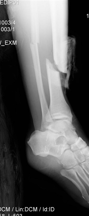
The A&E patient list wasn’t as exciting as I initially thought. However there were lots of post operative patients arriving from the wards and this gave me the opportunity to practice horizontal beam laterals (HBL). In the past I haven’t had much of an opportunity to have hands on practice for this type of examination. It is performed at Queen Margaret Hospital, using the air gap technique with the up-right bucky for trauma patients with a suspected neck of femur fracture. Here they perform their HBL without the air gap technique but the principals are the same. This examination is performed on patients who are post operative, trauma and some orthopaedic clinic reviews. I was given the chance to perform a few and through practice I found them not as difficult as I initially thought.

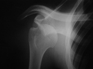
My other experience this week was performing an examination on a patient coming in through A&E with a suspected dislocated shoulder. The examination was quite difficult due to the patients’ pain, although the pain relief began to work and I was able to assist with the views needed. Once the examination was finished I was allowed to return with the patient to the resuscitation unit at A&E and observe while the doctor manipulated the shoulder back into position. It took a doctor and 2 nurses to perform the manipulation. While the doctor was pulling the patients arm down and away from the patients body, one nurse has a bed sheet placed around the top of the patients arm to pull the shoulder up, with the other nurse held the patient on the bed. After a few minutes of pulling, you were able to hear the patients arm slide back into position.

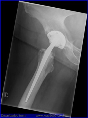
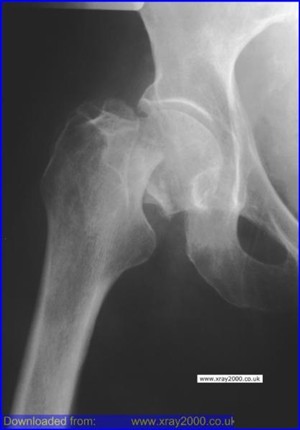
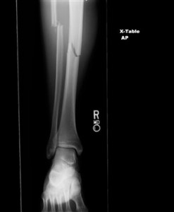
Attached to this piece of writing are images of fractured neck of femur and a hip replacement. A patient with a suspected fracture or a new replacement requires a HBL to show positioning of the fracture or replacement. There are also images of shoulder dislocations.


Week 6 Out of Hours Year 3

December 6th, 2009

This week’s placement was my out of hour’s week at Queen Margaret Hospital. I had been really looking forward to this week as I knew there were just two other radiographers working and I was hoping I would have a better chance of being involved with more of the trauma patients arriving in the department. This would give me the opportunity to deal with more challenging situations and the experience of adapting my technique.

Apart from the week having some quiet spells I had quite a few situations to deal with which required me to adapt my technique. I was also able to experience how the department works at night. I found this to be very exciting and I really loved my time in the department. I had advised the radiographers that I had been really looking forward to this week and was hoping to gain as much experience as possible in dealing with any trauma situations.

The first situation I was given was the chance to deal with was an elderly patient with a suspected fractured neck of femur (# NOF). There are a high number of elderly patients attending this department with this type of suspected injury due to the hospital being the main trauma centre. This type of injury has an increasing incidence with age thought to be due to a bone density loss and is more common in elderly females. This is usually due to a lack of oestrogen, which is common due to the menopause. Bone density loss can also been seen in patients taking a variety of medications, such as corticosteroids and thyroxine, with injury mostly due to only minor trauma. Classical features for this type of injury are the patients leg is usually shortened and externally rotated and pain on rotation and tenderness over the femoral neck.

The protocol for views needed at Queen Margaret for trauma situations dealing with suspected #NOF are an anterioposterior (AP) pelvis view and an air gap technique for the lateral (Lat) view. I had previously assisted in this situation before while on placement here, but had never been given the opportunity to perform these views. I had mixed emotions, excited, nervous and very cautious. I was comfortable with the radiographer I was working alongside knowing that they would allow me to proceed with the examination under supervision. I also knew they trusted me not to proceed with the exposure without them checking my positioning was correct first.

The AP pelvis was relatively straight forward although it can be difficult sometimes to see if all the correct anatomy will be on the cassette as patients tend not to lie in the middle of the trolley. I took my time and obtained an AP pelvis with all the relevant anatomy included for a #NOF, which was confirmed on viewing. I then positioned for the horizontal beam lateral (HBL), making sure everything was lined up as I thought it should be and then got the radiographer to check my positioning. I took the exposure and processed the image. On observing the image it was apparent that I could have centred down just a few more centimetres; however, the image showed clearly that the angulation of the head of femur was not in the correct position and the patient’s femur was displaced. We went on to repeat the HBL image which gave me the opportunity to repeat the examination and correct my positioning for a perfect image.

I was also able to visit resus on a few occasions, one occasion being for a gentleman who had fallen down the stairs and had injured has lower right leg. The patient had previous x-rays on his leg as he had a history of Osteomyelitis. This was my first time in resus in a long time so while the doctor was finishing his discussion with the patient the radiographer familiarised me again with the department. I had forgotten that they performed all the examinations with a mobile machine and had been expecting to be using a ceiling mounted tube. Realising this added to my nerves as I immediately thought the examinations were going to be difficult to achieve.

I had observed the patient while I entered the department and had observed that he was covered in blood and his leg was in an unnatural position. I had been advised a fractured tibia is usually associated with a fractured fibula and deformities are common. Deformities or angulation may be obvious on observation due to the foot being abnormally rotated. I obtained the request card to find out what views the doctor was requesting and advised the radiographer what views I thought would be the most appropriate projections.

The doctor had requested an AP and lateral views of the patient’s tibia and fibula from the knee down an AP foot and an AP hand. The most difficult part of the examinations was obtaining the distance for the lateral tib/fib, due to the room size. Moving the patient’s leg for positioning was relatively easy as he had self-anesthetised on alcohol.

I found both examinations very exciting to perform. They were new and challenging situations which I really enjoyed. On the whole the department was quiet due to my shifts being evenings and weekend. I felt I had more of an opportunity to relax and perform more challenging examinations without the pressure of a busy department, although I do feel one week is not enough. I would personally like more of an opportunity to do more out of hours work.

Attached to this piece of writing are images of fractured neck of femur and tib/fib.


Week 5 Endovascular Repair Year 3

November 27th, 2009

This was week 5 of placement and I was at Queen Margaret Hospital. It was my second day of placement and I was in a newly commissioned digital room. I was excited to be using the new equipment; however I was also very intimidated. Not only was there a new computer to learn to understand, I also found that I had to think differently about each examination. I had only just become comfortable with performing most of the examinations in rooms with a Computed Radiography (CR) system using cassettes, so the difference in environment threw me initially. I found in the beginning I was over thinking things especially when it came to performing extremity work. I was so used to using a cassette and thinking about how the image will look on the cassette and moving it to suit the patients’ position. I found myself thinking about how I could position the patient on the digital system so I could still have a good image.
It settled my nerves to know that even the radiographers I was working with were also a bit nervous using the new equipment. Later that afternoon there was a request of an abdomen examination. I had completed a few abdomen requests that day so didn’t have any problem in performing the examination until, that was, I had read the request in full. The request was for an AP, Lateral and Left and Right Anterior Oblique. I informed the radiographer that I wasn’t confident in performing the Lateral and the Left and Right anterior oblique as I had only read about there positioning techniques but had never saw them being performed.
The patient was in for a review due to having an endovascular aneurysm repair. I had never heard of this before and had to ask the radiographer what this was. She explained the patient had a stent placed in his aorta due to an aneurysm which was permanently dilated which is usually caused by a weakening in the vessel wall.
I performed the AP abdomen and the radiographer then performed the others while I observed. The images obtained were great, showing all sides of the stent for the patients’ consultant to review. While observing the images I could see the stent looked like wire mesh which extended from the lower part of the aorta to the upper parts of the common iliac artery, which look like a pair of metal trousers. I found this really interesting and have since done some research into understanding what is involved in endovascular aneurysm repair. I have attached some images and a website address, which shows a good animation of an aneurysm repair, to this piece of writing. This information helped increase my understanding of endovascular aneurysm repair.

http://www.stent-graft.com/

This website gives you lots of information and a good understanding of endovascular aneurysm repair, with animations of the procedure.


My personal development plan year 3

October 22nd, 2009

Current Situation

This is my plan of action for my PDP. I aim for development is in A & E radiography. I feel I haven’t had enough practice in adapting my technique in difficult situations. As a student you are not given the opportunity to perform and carry out examination in trauma situations or trolley patients. I feel I have the ability to logically carry out some of the examination I have come across but I very rarely been given the opportunity to carry them out.
I feel the experience in A & E would be of great benefit to me. Giving me the experience and build my confidence when performing them, hopefully making me a better radiographer.

Ideal Situation

My next block for clinical placement I have two ideal situations. First I am in Queen Margaret Hospital for 2 weeks, and 1 of them is my out of hours placement. This hospital is the main hospital for any trauma and I am hoping to be working with someone who will allow me to perform these examinations with supervision.
The following 2 weeks I am on my elective placement at Perth Royal Infirmary. This is a general placement and I have requested time within the department dealing with any trauma and A & E patients. I am hoping to have gained some experience and confidence from my time at Queen Margaret Hospital before proceeding to Perth Royal Infirmary. I am hoping these 2 weeks will be a great experience and of great benefit to me.

Steps to success

  • My 2 weeks placement at Queen Margaret Hospital.
    Learning to adapt my technique and keep calm in difficult situations. While performing as many trauma and trolley examination the 2 weeks I am there.
    • By performing as many examinations as possible on  trauma and trolley patients. by 04 December 2009
  • My 2 weeks at Perth Royal Infirmary.
    • By spending 2 weeks in A & E, and performing as many examinations as possible. Learning the best way to adapt techniques in difficult situations. by 18 December 2009

Overall Completion Date

18 December 2009

SWOT analysis

Strengths

My strength is that I friendly and good at chating to patients in a difficult situations. However this kind of situation is different to what I have ever experienced. I am also entering a new environment in Perth with new staff which I have never met.

Weaknesses

My biggest weakness is my confidence. Even though I know I am capable of most things that are asked of me I still question my ability. I am not sure if this is because we as students don’t get enough clinical time and are not always out on placement. My confidence begins to rise just as we are finishing our clinical placements.

Opportunities

I contacted Perth Royal Infirmary in the hope to get this elective placement. I spoke to a superintendent called Beryl Pritchard and explain what my goals were and what I was hoping to achievement while there.
I have a book which I will be referring to titled, Interpreting Trauma Radiographs. This book covers everything from, Pattern Recognition, Anatomy, Physiology and Pathology of the Skeletal System, Skeletal Trauma, Pelvic Fractures, Chest Trauma, and the Skull and Face.

Threats

Threats to this achievement are radiographers who don’t like students carrying out and performing examinations on patients in a trauma situation or in trolleys.
Another threat is the department is under staffed and I am asked to carry out general examinations to lower patient waiting times while the other staff deal with the trauma patients.

Supporting Resources

Books / journals

Interpreting Trauma Radiographs written by, J McConnell, R Eyres and J Nightingale.

Web links

http://www.wikiradiography.com/page/Pelvic+Trauma+Radiography
http://www.wikiradiography.com/page/Protocol+-+Trauma+Series

Other

Presentation

Reflection

I have just finished a 4 week placement comprising of 2 weeks and Queen Margaret’s Hospital, Dunfermline and 2 weeks at Perth Royal Infirmary and Perth. My experience at Queen Margaret hospital did not give me as much hands on experience as I was hoping for as my out of hours week was quiet. However I did get some opportunities to perform examinations where I had to adapt my technique in order to get good views.

I had the opportunity to practice and obtain a horizontal beam lateral (HBL) on a trauma patient who had a suspected neck of femur fracture. I also had to obtain views on a patient in resuscitation who had fallen down some stairs and fractured his lower leg.

Perth Royal Infirmary (PRI) was completely different from anywhere I had been previously. Being there presented many opportunities to refine and adapt my technique. The Accident and Emergency department is quite small and therefore there are generally not a lot of trauma cases passing through it. There was a big orthopaedic department which allowed me to see and perform techniques I have never observed before. This proved to be a great department to work and learn in, and I feel I have gained knowledge that is invaluable.

On the whole I feel that I have greatly benefitted from this placement. I learnt a large amount regarding adapting my technique for trauma and also views that I have previously read about but have never seen performed before. I think the main thing that I will take away from this placement is the fact that different trusts work in such different ways. I did find the number of changes quite over-whelming initially but by the end of my first week I was starting to adapt to the new routine and understand the system.

I feel writing this PDP has given me an opportunity to evaluate my own capabilities and has also highlighted my fears. By thinking of what aspects of my skill set I want to grow it has allowed me to set a realistic goal for my future development. I do feel this kind of experience is invaluable for me. My confidence did grow by the end of placement however I think this will diminish again the longer I am away from the department.


?>