/* Template Name: PageOfPosts */ Angie's CPD » Reflectives

Week 6 Year 4

January 5th, 2011

04 January 2011

The start of this week’s placement at Victoria hospital was fairly busy and I was able to achieve plenty of hands on experience, however by the end of the week the department was having technical problems with their equipment. I was mainly in one room for the whole of the week. On the Monday the room where I was due to be working had a problem with the table top. So we spent our time going through the appropriate examinations which were able to be performed in the room to try and keep the patients waiting time to a minimal. The work load was stream-lined to make full use of the room. By late afternoon the engineer arrived to fix the table top. Tuesday the room was up and running and the department was busy. For the next two days I really enjoyed my time in the department. I was working along side a very experienced radiographer who gave me all her attention and explained some very good techniques. One of these techniques was for a supine cervical spine examination. I have only once performed this examination and found the concept to be quite difficult to grasp, however the radiographer took the time to explain and demonstrate the procedure to allow a better understanding of the technique. Another examination she explained was in the ball catchers’ examination for rheumatology, she explained she was taught to lay the backs of the hands on the cassette and get the patient to curl their fingers slightly to look like they were going to catch something. She explained it was a necessary part of the examination was to demonstrate all joint spaces for evaluation. She explained although my technique was not wrong I could demonstrate the joint spaces better if I used her technique. I have since had the opportunity to perform the ball catchers’ examination however I have not as yet been able to perform and try the supine C-spine examination. I have previously worked with this radiographer and on every occasion she has always given me her full attention and co-operation and what I think to be valuable knowledge which I can use throughout my training.
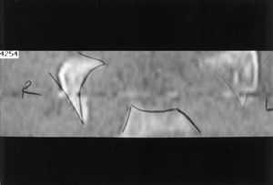
During the week I performed an examination on a patient who was referred to the department form her General Practitioner (GP) due to onset of pain with an inability to weight bear.
I went to the waiting room and called the patient and provided her with a gown and advised her to change for the exam. Once she had changed I advised the patient I was a student in the department and asked her consent to perform the examination. While she was entering the room she seemed uncomfortable but did not complain of any pain. Once the examination was done it became obvious from the x-ray that the patient had a fracture of both the superior and inferior pubic ramus. The procedure then was to refer the patient round to the Accident and Emergency department. Fractures of the pelvis can be caused by a direct blow, e.g. direct fall, which may cause damage to the bladder or urethra, or major blood vessels.

According to Dutton (2004), the superior pubic ramus is the most commonly fractured of the pubic rami and account for more than 70% of all pelvic fractures. Signs of a pubic rami fracture are the gradual onset of pain in the groin which is aggravated by weight bearing, walking or abduction of the thigh.
According to Misra and Holmes (2004), a simple pubic rami fracture can often be discharged with analgesia following assessment of their home situation. While unstable fractures require adequate fluid resuscitation and early fixation. Most external fixation can be treated in an A&E department by experienced personnel and considerations for potential injuries, such as urethal or rectal disruption always have to be considered.
Attached to this piece of writing are images of fractured superior and inferior pubic ramus and a Medscape document on pelvic anatomy and classifications of pelvic fractures.
Dutton, M. 2004. Orthopaedic Examination, Evaluation, and Intervention. McGraw-Hill Professional Publishing, New York, New York, USA.
Misra, R. R. and Holmes, E. J. 2004. A-Z of Emergency Radiology. Cambridge University Press, West Nyack, NY, USA
http://sinoemedicalassociation.org/orthopedicsurgery/pelvicfractures.html

http://e-radiography.net/radiology/acetabular%20fractures.pdf


Week 5 Year 4

January 4th, 2011

This week I was at Victoria Hospital. During my time this week I was assisting in Endoscopic retrograde cholangiopancreatography (ERCP). There was a full morning list and we were using a new room. I had previously screened ERCP patients in this hospital, however this was new equipment that I was being instructed on.
ERCPs are performed on patients who suffering from gallstones or experiencing problems such as jaundice. Ducts in the biliary system drain bile from the liver and pancreas. The biliary ducts and the pancreatic ducts join just before they drain into the upper bowel. This drainage opening is called the papilla and is surrounded by a circular muscle, called the sphincter of Oddi.
One patient due to have her gallstones removed had to have a biliary sphincterotomy. This is where the surgeon has to cut the muscle which surrounds the opening of the duct. This cut is made using a specialised catheter which has an electric current running through it. The surgeon was able to see stones in the gall bladder but was unable to remove them without performing the sphincterotomy. Once the sphincterotomy had been performed to enlarge the opening of the bile duct, the stones were able to be pulled from the duct into the bowel using a balloon attached to the catheter. Once the stones were removed the patient was experiencing a little bleeding. The surgeon then explained to me he was going to inject adrenaline around the site of the cut to try and minimise the bleeding. Epinephrine commonly referred to as adrenaline is a naturally produced hormone within the body, secreted by the medulla of the adrenal glands. Epinephrine, is used to contract the blood vessels around the site of the cut. Epinephrine is a hormone and a neurotransmitter. It can be used to increase the heart rate, contract blood vessels, and dilate air passages and participates in the fight or flight response of the sympathetic nervous system. Epinephrine is added to injectable forms of local anesthetics such as lidocaine as a vasoconstrictor.
Another procedure which I performed was the screening of patients who had undergone a procedure called adiana. This procedure is a minimally invasive procedure that permanently prevents pregnancy. It works by stimulating your body’s own tissue to grow in and around tiny soft inserts that are placed inside your fallopian tubes. This is a simple procedure with a quick recovery and leaves nothing in the uterus that might limit future gynecologic procedures. It is performed by inserting a catheter into the cervix and into the uterus. This catheter delivers a low level radiofrequency (energy that generates heat to create a superficial lesion) to a small section of each fallopian tube. A tiny soft insert the size of a grain of rice is placed in each of your fallopian tubes where the radiofrequency is applied. This allows for new tissue to grow in and around the adiana inserts, eventually blocking your fallopian tubes. Patients are then sent for a hysterosalpingogram (HSG) to confirm that the tubes have been fully blocked. This test is performed to ensure that the procedure has been successful.
Attached to this piece of writing are images of both procedures.

Endoscopic Treatment for Bleeding Peptic Ulcers. 2010. Available at: http://sunzi.lib.hku.hk/hkjo/view/23/2300709.pdf [Accessed October 30 2010].
ERCP. 2010. Available at:http://emedicine.medscape.com/article/365698-imaging [Acessed October 30 2010].
Sphinterotome. 2010. Avaliable at; http://www.top5plus5.com/Procedures_files/THERAPEUTIC%20ENDOSCOPY.htm [Acessed October 30 2010].
Colonoscopy. 2011. Available at:http://www.colonoscopy-exam.info/coe/Portals/0/proc_images/procedure_images/photo14.jpg [Accessed October 30 2010].
ERCP. 2011. [online image] Avaiable at: http://www.google.co.uk/imgres?imgurl=http://www.pregnantagain.com/img [Accessed October 30 2011].
ERCP. 2011. [online image] Avaiable at:http://www.google.co.uk/imgres?imgurl=http://journals.prous.com/journals/ [Accessed October 30 2011].


Adrenaline used in surgery


Week 4 Year 3

January 4th, 2011

SkullThis was week 4 placement and I was now at the Western General hospital. I always like working at the Western General hospital as the staff are very willing to teach and there is lots of opportunity to get of a plenty of hands on experience.
The whole week was very busy and I gained a lot of valuable experience in all aspects of the job. One examination that made me nervous was regarding a request for a patient that was due to have a Magnetic Resonance Imaging (MRI) scan. The request was to perform an orbits examination prior to him having his MRI scan due to the pre-MRI safety check, performed by the radiographer, highlighting that this patient had previously worked with metal.
I had performed an orbits exam in the past using a skull unit but was nervous about attempting it using an up-right bucky. This was due to my lack of experience in both using this equipment and being inexperienced at performing the an orbit exam. I discussed how I was going to attempt the examination with the radiographer and we agreed I would position the patient and she would check my positioning before I proceeded to complete the exam.
The basic orbits (OM) examination as stated by Carver and Carver (2006) is performed by;
• The patient seated in front of the image receptor, (this should help to ensure some stability to the patients’ posture and balance).
• The chin is placed in contact with the midline of the image receptor and the chin position adjusted until the Orbitalmeatal Baseline (OMBL) has been raised 30˚ from the horizontal plane.
• The Medial Sagital Plane (MSP) is perpendicular to the image receptor, which is assessed by checking that the External Auditory Meatus (EAMs) or lateral margins are equidistant from the image receptor.
• Centre above the External Occipital Protuberance (EOP) to emerge level with the middle of the orbits.
• Collimate to include the orbits and maxillary sinuses.

The examination was actually straight forward and once I had had time to reflect on it then it was relatively easy. The initial fear came from trying to adapt an exam that I had previously only performed using a skull unit to performing the exam on an upright bucky. On reflection, the examination was actually easier using an upright bucky than it was using the skull unit as there was less to think about in terms of positioning the cassette and the positioning angle of the skull unit.
When positioning for any examination of the skull or facial bones it is important the patient is perfectly straight with no rotation or tilt. Common errors in positioning are caused when the MSP is not parallel to the cassette and the interpupilary line is not at 90Ëš to the film. Positioning errors can be reduced by using the eyes rather than the nose as positioning aids as it is recognised that the human body is not always symmetrical.
I have very little experience in head/skull radiography and still find it intimidating and challenging especially when trying to interpret skull images. I feel from a students’ perspective it is difficult to fully understand positioning techniques from the text books. I feel observing examinations being performed and having hands on experience in all aspects of these examinations is extremely important in all types of skull examinations. However demand for plain film imaging of the head/skull has been reduced greatly due the introduction of Computed Tomography (CT) and Magnetic Resonance imaging MRI.

Attached to this piece of writing are images of a skull unit and images and diagrams of the skull taken from; http://www.e-radiography.net.

Clark, K.C. 2005. Clarks positioning in radiography. 12th ed. London: Arnold.


Week 3 Year 4

January 4th, 2011

This was my third week at Crosshouse hospital and it was my first proper opportunity to get full time patient contact due to the previous two weeks being CT and in wards and theatres.

My skills felt quite rusty due to having no proper hands on experience for such a long period of time and I felt nervous. Protocols are also different at Crosshouse but I knew this from the last placement I had there. My first two days were mentally exhausting from double checking everything but I found it really exciting.

It was quite a slow week with not many clinics however during one orthopaedic clinic I encountered an interesting problem. The request was for a right sternoclavicular joint projection. The patient had sustained an injury which had caused subluxtion of the right sternoclavicular joint and was attending the department for a review examination from the orthopaedic consultant.

I had received a few requests from this particular orthopaedic consultant that I had not encountered before and had only read about in the positioning books which I found very interesting. These requests were for lateral scapula, sternoclavicular joints and acromioclavicular joints.

I talked the examination through with one of the radiographers; however she was also not confident in performing this exam. We then got the departments positioning book out for reference but then requested the help of a more experienced radiographer. It was acknowledged this was a rare request and due to the lack of familiarity of this particular position this examination was performed by the member of staff that felt most confident.

The positioning book dictated that we use the Kurzbauer method which states it is an unobstructed lateral projection of the sterno-clavicular articulation. This describes that the patient lies on the affected side with the arm they are lying on next to their head. Using a vertical central ray, directed 15 degrees caudally and centered to the lower most sterno-clavicular articulation. This technique was used but modified by the radiographer so the patient was positioned standing with the affected side against the upright bucky.

While observing this technique it seemed straight forward and relatively easy to perform. Once the image was obtained I found it difficult to interpret and had to get the radiographer to explain what we were looking at. After interpretation of the image it was difficult to see if any changes had occurred, as there were no previous images to compare them against. It is difficult to gain experience in these examinations since they are so rare. There was only one more request for a sternoclavicular joint that day and a different radiographer performed it while being observed.

Throughout the week I started to regain my confidence slowly with the examinations that are requested more frequently. However it took some time as I struggled to remember some protocols and also had to remember how to interact with the patients (radiology information system) RIS system.

I really enjoyed the experience of the various new referrals which had been requested while this clinic was on and I am really looking forward to tackling them in the future.

Attached to this piece of writing is an orthopaedic booklet on Sternoclavicular Joint Separation and images of the shoulder and the AC joint, and also a CT scan of a patient’s right and left sternoclavicular joints. Relative to the sternum, the left medial clavicle demonstrates 4 mm of superior subluxation. There is no detectable superior subluxation of the right medial clavicle.

Pearsall, A.W. and Russell, G.V. 2000. Ipsilateral Clavicle Fracture, Sternoclavicular Joint Subluxation, and Long Thoracic Nerve Injury: An Unusual Constellation of Injuries Sustained During Wrestling. The American Journal of sports medicine 28 (6) February, pp.904-8. Available at:http://ajs.sagepub.com/content/28/6/904.full.pdf+html [Acessed October 20 2010].

Eorthopod. 2010. Sternoclavicular problems. [online] Available at: http://www.eorthopod.com/content/sternoclavicular-joint-problems [Accessed October 20 2010].

Shoulder. 2010. Available at:http://www.projectswole.com/weight-training/the-top-5-best-shoulder-exercises/ [Accessed October 20 2010].


A Patient Guide eOrthopod


?>