Week 8 Year 4
Week 8
This week I was out of hours. It was a quiet week; however there were a few mobile exams and a few occasions where I had to adapt my technique in order to achieve a good result. A few of these examinations being chest x-rays on patients who were unable to co-operate due to either being unconscious or medicated. I also had a few wrist x-rays pre and post manipulation where I had to adapt my technique to perform them.
Patient Positioning
The optimal positioning for obtaining a chest radiography is with the patient postero-anterior (PA) and erect (Clarks 2005). However this is not always possible, due to patient conditions some radiographs have to be obtained antero-posterior (AP) with the patient in a sitting or semi-recumbent or supine position. Patient positioning has a significant influence in the appearance of air or fluid, and blood vessels within the chest: Air usually rises to the highest point within the chest cavity. Pathologies such as a pneumothorax are mostly seen in the apex of the lung in an erect chest x-ray. When the patient is supine, the highest point in the chest lies alongside the heart and mediastinum. According to medscape 2010, a pneumothorax may cause increased lucency adjacent to these structures, which appear to have a better-defined outline than normal, often with no lung edge visible.
On an erect film any fluid lying in the lungs usually collects at the lung bases and appears as cloudy or opaque and obscures adjacent structures. Fluid levels in the lung are higher on the lateral wall of a chest image and lower at the mediastinum. When performing a supine examination, any fluid in the lungs would lie along the posterior chest wall.
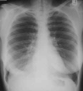
A technical problem encountered by student radiographers while performing mobile chest x-rays are Lordotic AP films sometimes referred to as an apical lordotic view. A lordotic image is produced when the tube angle is not in the correct position. One of the first things you are taught when performing mobile chest exams is to always try to sit the patient as up-right as possible while giving consideration to the patients condition. Although you are taught in lectures about how a lordotic image is achieved, it sounds easy to avoid. This has been something I have had to consider a lot on placement this week.
A technique shown to me while on placement which should eliminate the chance of producing a lordotic image is to angle the tube perpendicular to the sternum so it runs parallel to the sternum and then over compensate and increase the angle down slightly more. This is now a technique which I have managed to try and has worked however I have only tried it once so I am looking forward to trying it a few times to evaluate if it works every time.
In a lordotic film the clavicles are projected higher than normal over the lung apices and the posterior and anterior ribs appear flattened. Attached to this piece of writing is a lordotic image for demonstration.
Carver, E. and Carver, B. 2006. Medical imaging: techniques, reflection and evaluation. Edinburgh: Churchill Livingston Elsevier.
Clark, K.C. 2005. Clarks positioning in radiography. 12th ed. London: Arnold.
Martensen, K.M. 2006. Radiographic image analysis. St Louis: Elservier.
http://bloggingradiography.blogspot.com/2007/07/lo
 Print This Post
