Week 7 Year 2
Journal
This week on placement I had a number of patients from an oncology clinic for follow up chest x-rays. These patients were all referred due to having conditions called seminoma and teratoma. Both these conditions are cellular cancers which
started, in these cases, in the testes. Seminoma is a radiosensitive malignant neoplasm of the testis, and teratoma is a germ cell tumour composed of multiple cell types derived from one or more of the 3 germ layers.
According to emedicine, 3% of testicular teratomas are known to metastasize in adults and adolescents. There are two types of teratomas, mature and immature. Mature teratomas are usually found in women and are usually found to be benign, whereas the immature teratomas are usually found to be malignant and more commonly found in males. Teratomas are thought to be congenital, but are often not diagnosed until later in life.
The patients had all been referred for follow up chest x-rays by their consultant to check for any metastases in the lungs. I had never heard of any of these diseases before and looking over the request cards I noticed all the patients were all young men, between the ages of twenty five and thirty five. After some research into these diseases I discovered they were both cellular cancers, beginning in the embryonic stage. Once discovered, these diseases are monitored carefully as there is the possibility for them to metastasize.
All patients x-rayed had no obvious signs of metastases and were due to see their consultant after their x-ray. Attached to this piece of writing are images and a website I used to research these diseases. Throughout my research I found out both male and females can have this condition, however in this case, all patients were male.
Both seminoma and teratmoas can be found in different parts of the body. According to emedicine, the most common location is sacrococcygeal. As they arise from totipotential cells, they are encountered commonly in the gonads.
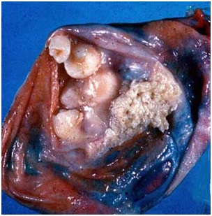
The most common location being the ovary, and occurring less frequently in the testes. Occasionally teratomas occur in midline embryonic cell rests and can be mediastial, retroperitoneal, cervical, and intracranial. Cells differentiate along various germ lines, essentially recapitulating any tissue of the body. Examples include hair, teeth, fat, skin, muscle, and endocrine tissue.
Testicular cancer treatment involves radical surgical. This surgery involves the removal of the testicle. As part of the diagnosis of testicular teratoma, tissue diagnosis and distinction from other forms of testicular cancer is important in subsequent management. Regional lymph nodes may also be sampled or removed during surgery. Teratoma of the testicle is relatively resistant to radiotherapy but responds well to chemotherapy as an additional testicular cancer treatment to surgery or as a primary treatment in advanced disease.
Following the surgery or chemotherapy the patient will be closely followed to detect any recurrences early. Follow up includes self-testicular examination, measurement of tumour markers in the blood, serial chest x-ray and regular
abdominal and thoracic CT scanning.
 Print This Post